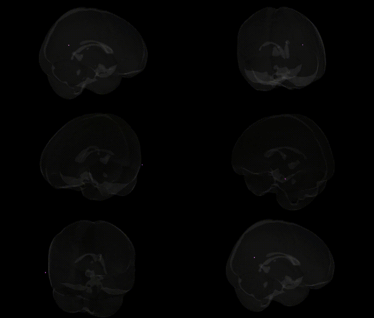
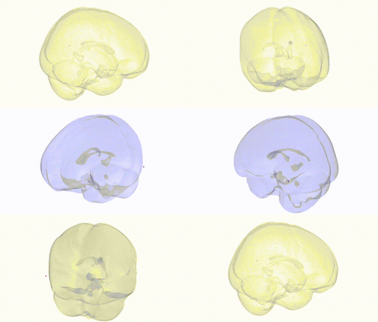
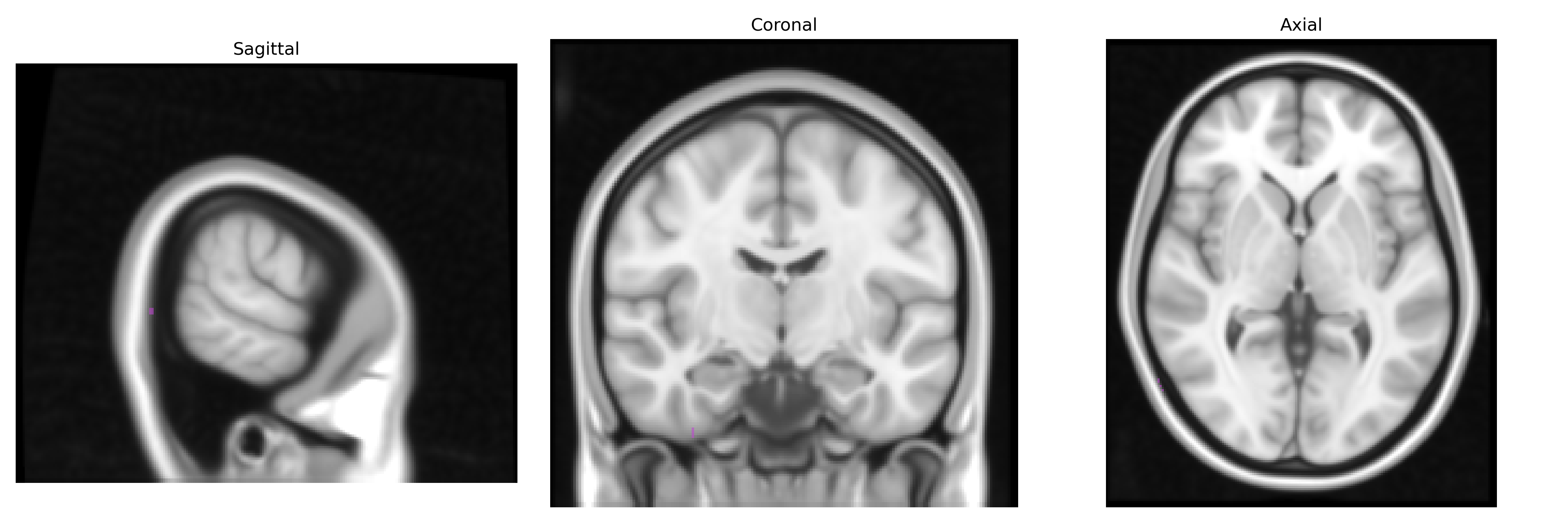
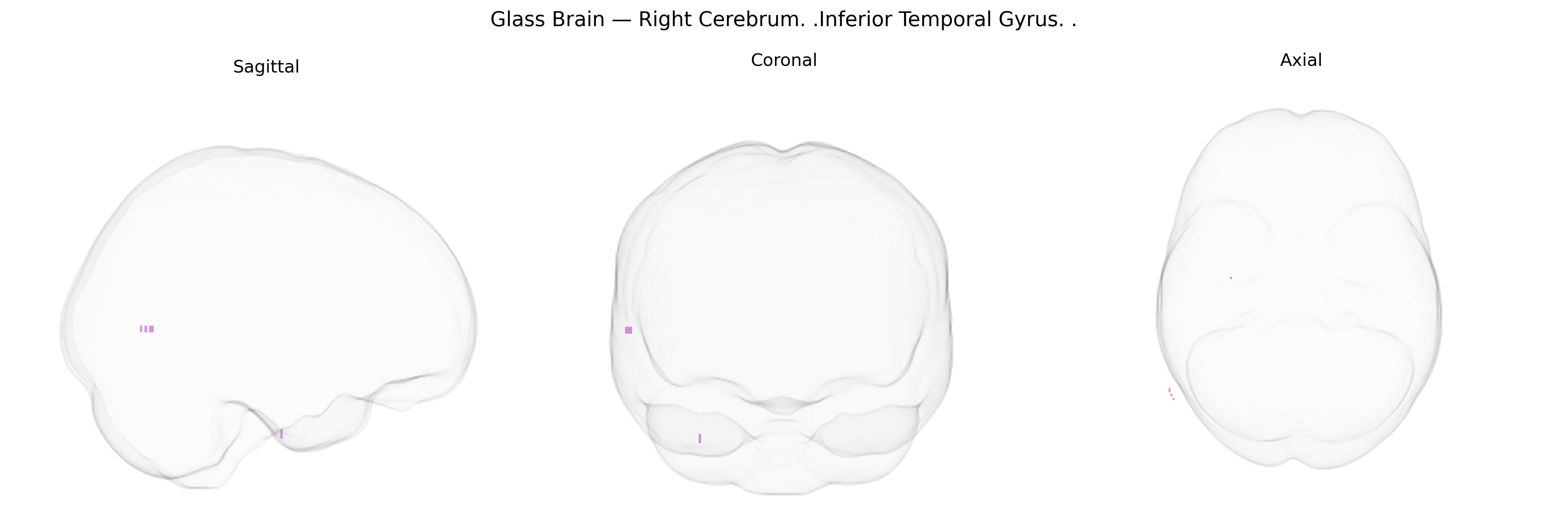
The bilateral Right Cerebrum Inferior Temporal Gyrus is a cortical region located on the ventrolateral aspect of the temporal lobe, extending along the inferior surface between the middle temporal gyrus laterally and the fusiform gyrus medially. It is primarily associated with high-level visual processing, including object recognition, complex shape and pattern analysis, and aspects of visual memory, with functional integration into the ventral visual stream. This gyrus participates in semantic processing and may contribute to language-related functions, especially in the dominant hemisphere, while also interfacing with limbic and association cortices for multimodal integration. Lesions in the inferior temporal gyrus can lead to deficits in visual object recognition (agnosia) and disturbances in semantic knowledge. There is no direct link, but a related structure is the Inferior temporal gyrus.
The bilateral right inferior temporal gyrus (ITG), a key high-level visual and semantic processing region, has been repeatedly implicated in genetic studies of cognition and neuropsychiatric disease, though often under broader temporal or ventral visual cortex labels rather than Talairach-specific coordinates. GWAS of cortical morphology (e.g., ENIGMA and UK Biobank cohorts) have identified common variants in genes involved in neurodevelopment (such as KIAA0586, MAPT, and nearby loci on chromosomes 17q and 6p) associated with temporal cortical thickness, surface area, and gyrification, which include the inferior temporal territory; these structural metrics, in turn, are genetically correlated with general cognitive ability, educational attainment, and risk for disorders like schizophrenia and major depressive disorder. ITG activation and volume show heritability in twin studies and have been linked via imaging genetics to variants in synaptic and plasticity-related genes (including BDNF Val66Met, COMT, and DISC1), influencing semantic memory, face and object recognition, and reading-related functions. In autism spectrum disorders, rare and common variants affecting synaptic scaffolding (e.g., SHANK3, NRXN1) and chromatin remodeling (e.g., CHD8) have been associated with atypical activation and connectivity in temporal association cortex that encompasses the ITG, particularly for social and language-related tasks. Alzheimer’s disease GWAS loci such as APOE, CLU, and PICALM show downstream effects on temporal lobe atrophy and amyloid burden in ventral temporal regions, including the inferior temporal cortex, with ITG involvement in early semantic memory decline. Additional GWAS and candidate gene studies of reading disability and dyslexia (e.g., DCDC2, KIAA0319, ROBO1) have reported structural and functional alterations in left-lateralized inferior temporal and adjacent fusiform regions, with some evidence of homologous right ITG involvement in visual word processing networks. Overall, genetic influences on neurodevelopment, synaptic function, and neurodegeneration converge on temporal association cortices, and large-scale imaging-genetic consortia support polygenic contributions to right ITG structure and function that modulate vulnerability to cognitive and psychiatric phenotypes, even though few loci are uniquely mapped to this specific Talairach-defined region.
Overview generated by GPT-4o (2026).
Region ID: 47
Hemisphere: bilateral
Atlas: Talairach labels 1mm
Full Quality Version: Download MP4
Full Quality Version: Download MP4
Wali Sidiqyar*, Gaurav Rudravaram*, Elyssa M. McMaster, Trent M. Schwartz, Adam M. Saunders, Kurt G. Schilling, Bennett A. Landman "Introducing SPINS: A Shared Public Visualization Library of Neuroanatomical Structures." Medical Imaging with Deep Learning- short paper
This resource is licensed under CC0 1.0 Universal (Public Domain).