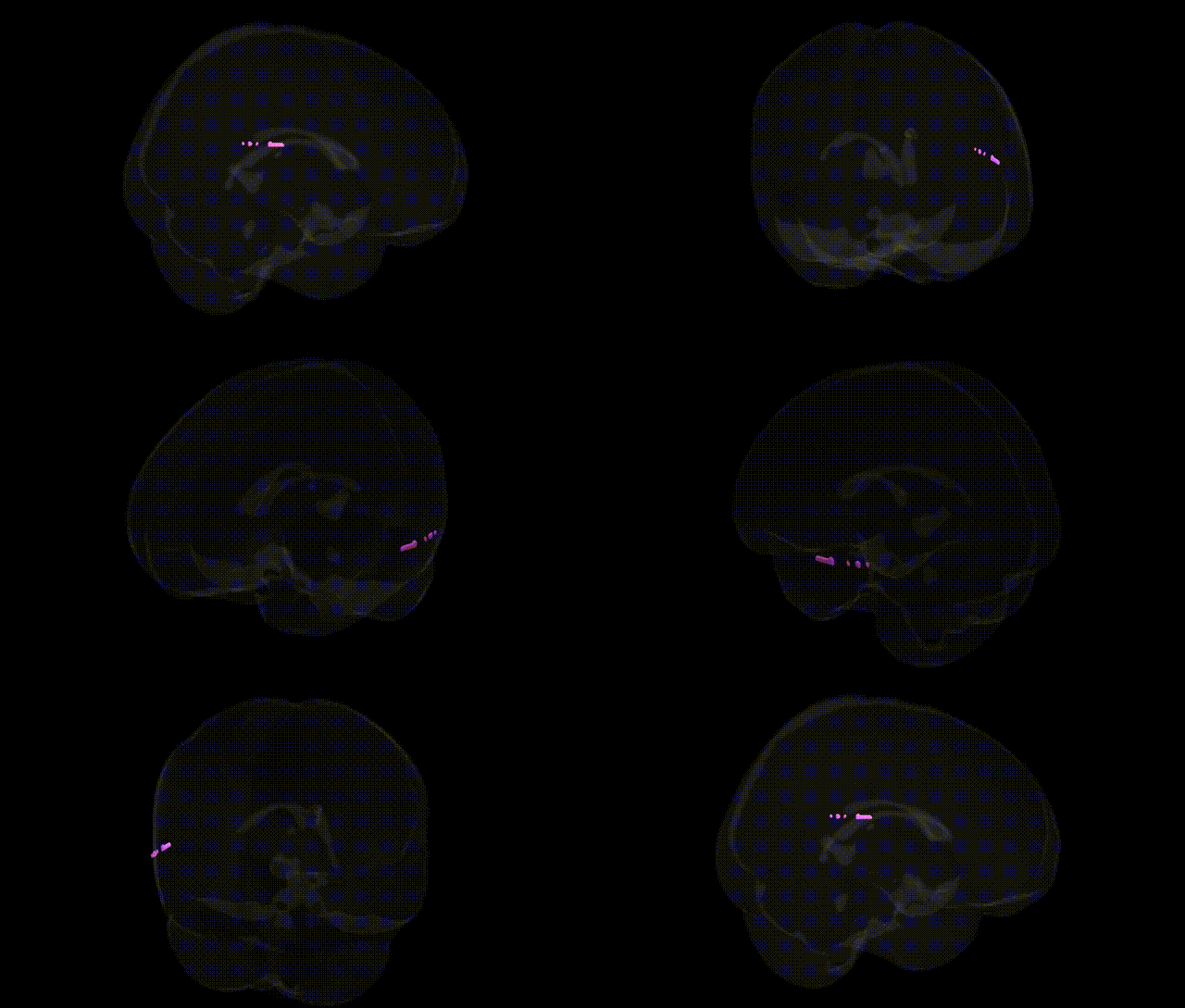
The bilateral Right Cerebrum Middle Temporal Gyrus is a cortical region of the temporal lobe extending along the lateral aspect of the right hemisphere, situated between the superior and inferior temporal gyri and spanning anterior to posterior portions of the temporal lobe. In the Talairach 1 mm Atlas, it corresponds to portions of Brodmann areas commonly associated with auditory association, semantic processing, lexical retrieval, and aspects of social cognition, including interpretation of biological motion and theory-of-mind–related cues. This gyrus participates in multimodal integration of auditory, visual, and linguistic information, contributes to comprehension of word meaning and sentence-level semantics, and interfaces with wider temporal–parietal and frontal networks supporting language and memory. Functional lateralization in the right hemisphere emphasizes roles in prosody, discourse-level interpretation, and nonverbal social signals. A related article is Middle temporal gyrus.
The bilateral right middle temporal gyrus (MTG) has been implicated in multiple genetic and GWAS findings, particularly in relation to language, social cognition, and neuropsychiatric disorders. Imaging genetics studies from large consortia such as ENIGMA and UK Biobank have identified common variants in genes involved in synaptic function, neurodevelopment, and axon guidance (for example, in pathways including NMDA receptor signaling, cell adhesion molecules, and Wnt signaling) that are associated with MTG cortical thickness, surface area, and volume. GWAS of schizophrenia, autism spectrum disorder, major depressive disorder, and bipolar disorder frequently report structural or functional abnormalities in the right MTG in carriers of polygenic risk, and polygenic risk scores for these disorders correlate with altered MTG morphology and connectivity. Variants in FOXP2 and related language-network genes have been linked indirectly to right MTG activation during speech and semantic processing, while Alzheimer’s disease and frontotemporal dementia risk loci (including those near APOE, CLU, and GRN) are associated with atrophy patterns that prominently involve the temporal neocortex, including the MTG. Additionally, GWAS of cognitive traits (such as general intelligence, educational attainment, and reading-related abilities) and social cognition show genetic correlations with right temporal-lobe structural measures, suggesting that shared polygenic architectures shape both MTG anatomy and higher-order cognitive functions.
Overview generated by GPT-4o (2026).
Region ID: 563
Hemisphere: bilateral
Atlas: Talairach labels 1mm
Full Quality Version: Download MP4
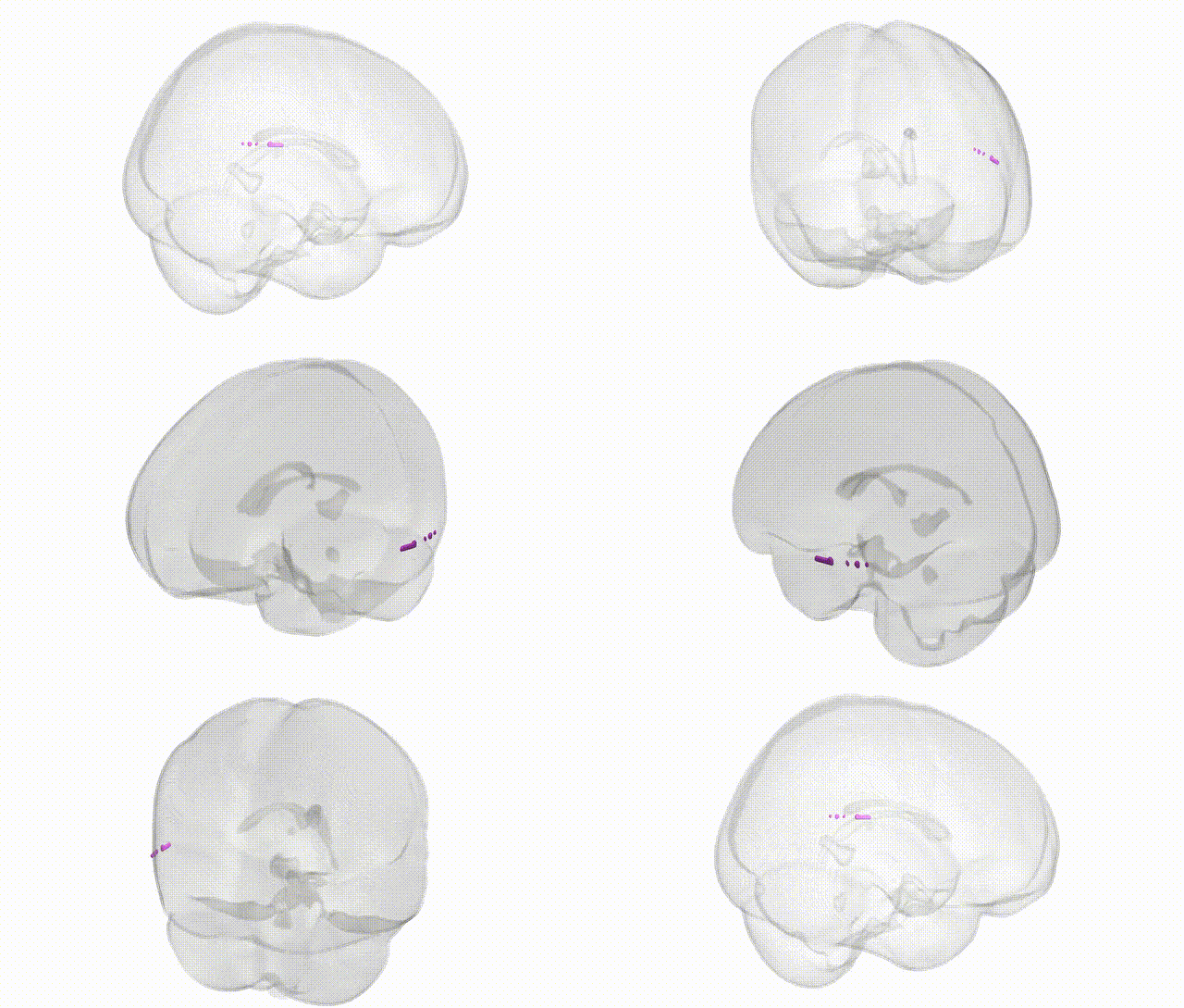
Full Quality Version: Download MP4
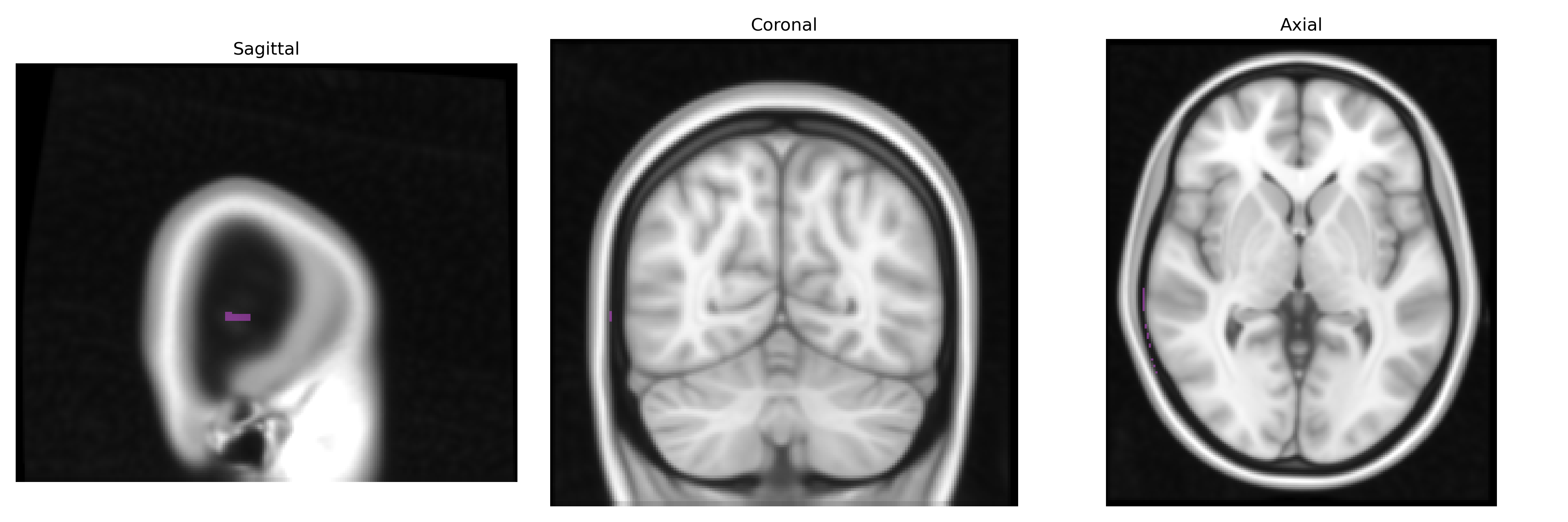
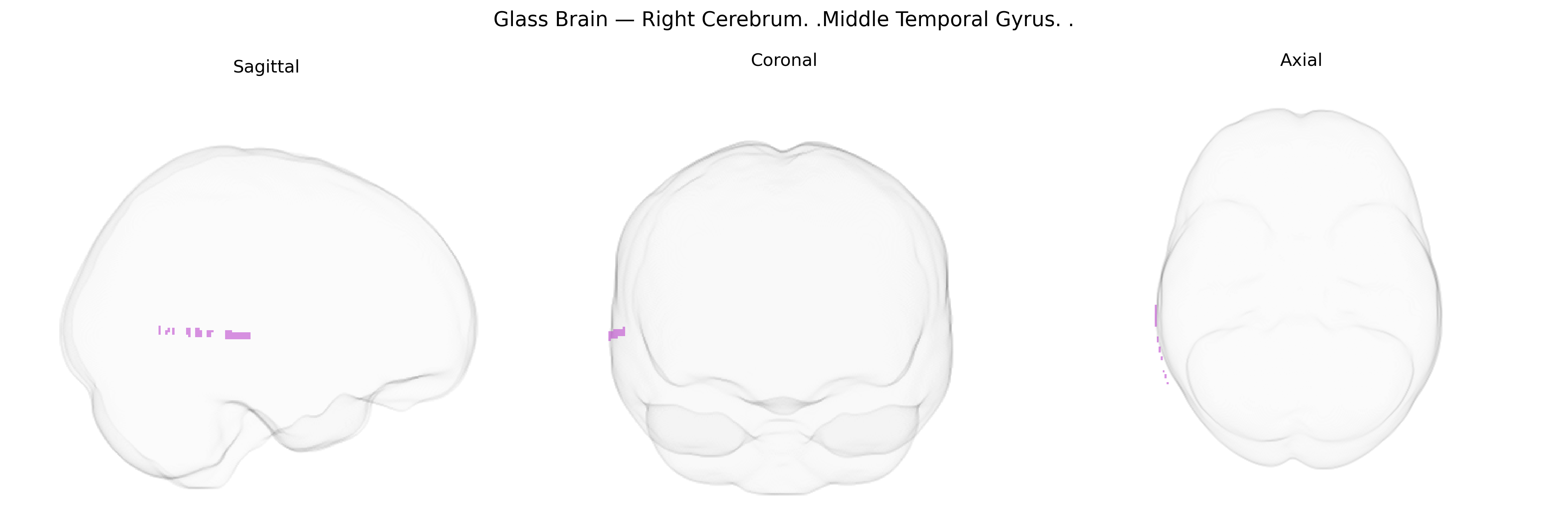
Wali Sidiqyar*, Gaurav Rudravaram*, Elyssa M. McMaster, Trent M. Schwartz, Adam M. Saunders, Kurt G. Schilling, Bennett A. Landman "Introducing SPINS: A Shared Public Visualization Library of Neuroanatomical Structures." Medical Imaging with Deep Learning- short paper
This resource is licensed under CC0 1.0 Universal (Public Domain).