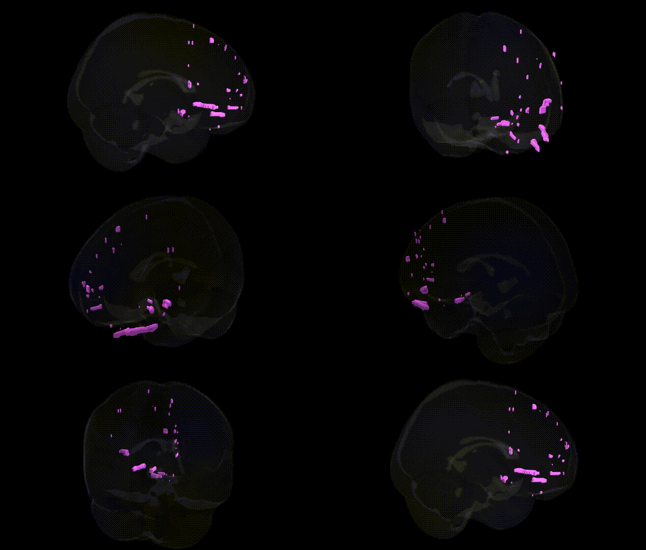
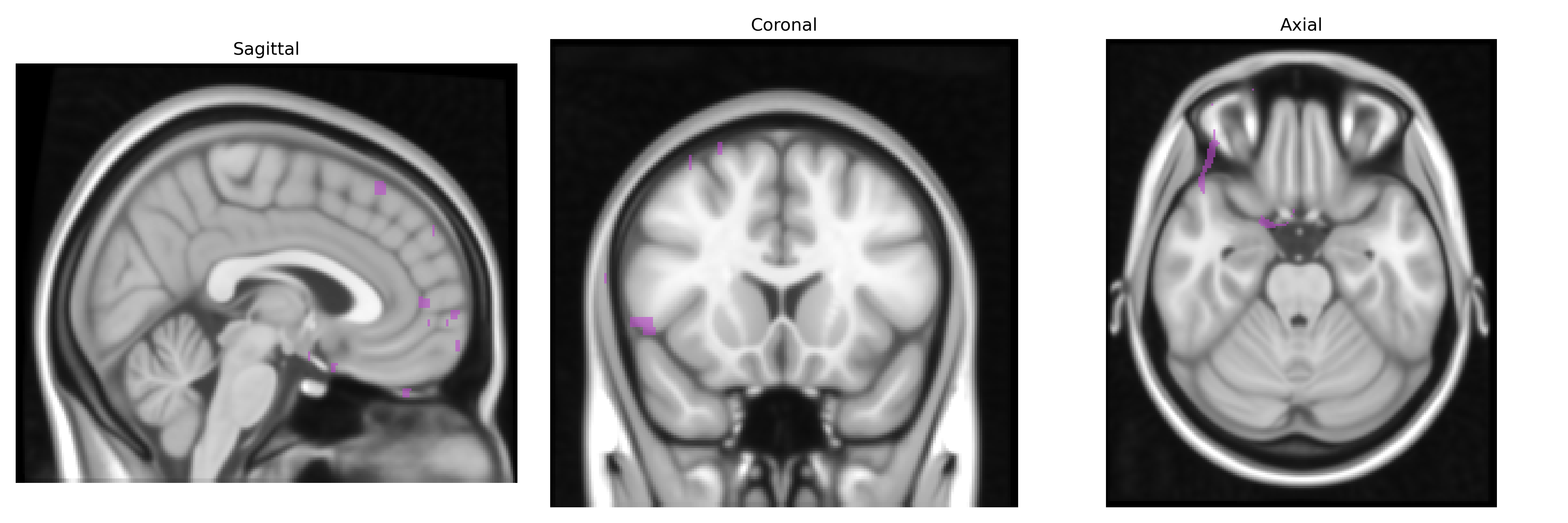
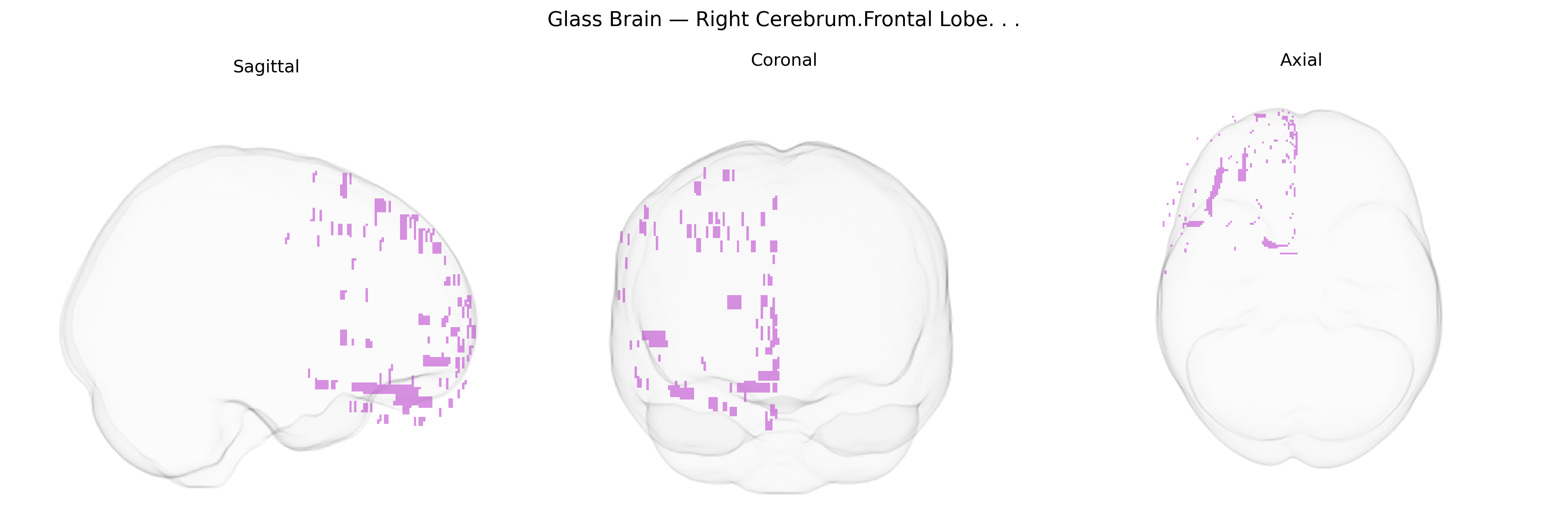
The bilateral Right Cerebrum.Frontal Lobe, as defined in the Talairach 1 mm atlas, encompasses the right frontal cortical mantle anterior to the central sulcus, including premotor, supplementary motor, and prefrontal territories involved in higher-order motor planning, executive function, working memory, attention, and aspects of social cognition and affect regulation. This region integrates multimodal sensory inputs with subcortical signals to modulate goal-directed behavior, decision-making, and inhibitory control, while also contributing to language-related and nonverbal communicative functions, particularly in prosody and pragmatic aspects of speech. Anatomically, the right frontal lobe overlies the anterior cranial fossa and is supplied predominantly by branches of the middle and anterior cerebral arteries, with extensive reciprocal connectivity to parietal, temporal, limbic, and basal ganglia structures, supporting its role in coordinating complex cognitive and behavioral repertoires. Frontal lobe
The bilateral right frontal lobe, as defined in the Talairach 1 mm atlas, is implicated in a wide range of genetic associations linked to cognition, behavior, and neuropsychiatric disorders. Large-scale GWAS of brain imaging phenotypes (e.g., ENIGMA, UK Biobank) have identified common variants near genes such as HMGA2, FOXO3, KIAA0586, and others that influence global and regional frontal lobe volume, cortical thickness, and surface area. Polygenic risk scores for schizophrenia, bipolar disorder, major depressive disorder, ADHD, and autism spectrum disorder consistently associate with structural and functional variation in frontal regions, including right dorsolateral and ventrolateral prefrontal cortex, reflecting shared neurodevelopmental pathways across these conditions. Variants in genes linked to synaptic function and neurodevelopment (e.g., CACNA1C, GRIN2A/B, CNTNAP2, DISC1) have been related to altered frontal activation and connectivity in tasks tapping executive function, working memory, and inhibitory control, while APOE ε4 and other Alzheimer’s disease risk loci have been associated with right frontal atrophy and hypometabolism in aging and prodromal dementia. Additional GWAS have connected frontal morphology and activity to general intelligence, educational attainment, risk-taking and impulsivity, substance use, and personality traits (such as neuroticism and extraversion), indicating that genetically driven variation in this region contributes to both disease vulnerability and normal behavioral diversity.
Overview generated by GPT-4o (2026).
Region ID: 108
Hemisphere: bilateral
Atlas: Talairach labels 1mm
Full Quality Version: Download MP4
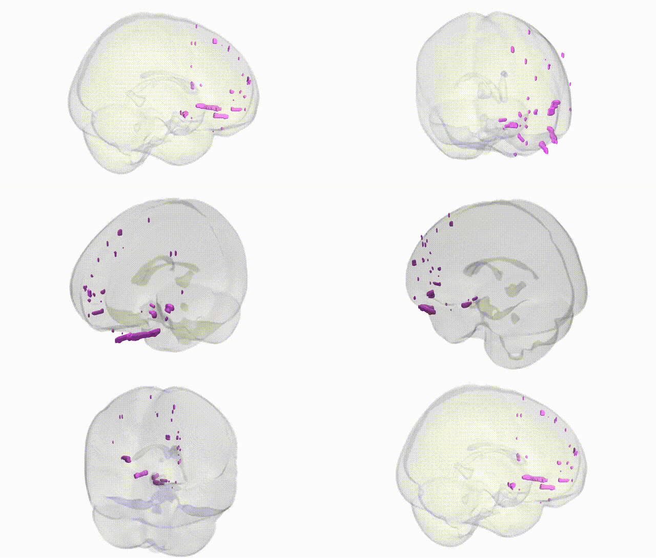
Full Quality Version: Download MP4
Wali Sidiqyar*, Gaurav Rudravaram*, Elyssa M. McMaster, Trent M. Schwartz, Adam M. Saunders, Kurt G. Schilling, Bennett A. Landman "Introducing SPINS: A Shared Public Visualization Library of Neuroanatomical Structures." Medical Imaging with Deep Learning- short paper
This resource is licensed under CC0 1.0 Universal (Public Domain).