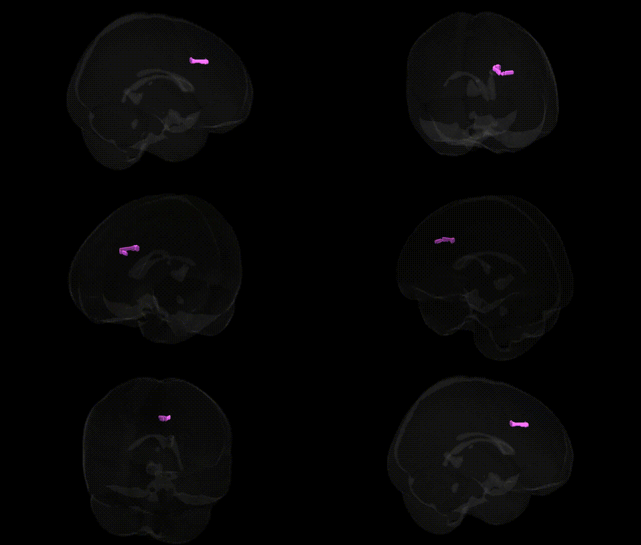
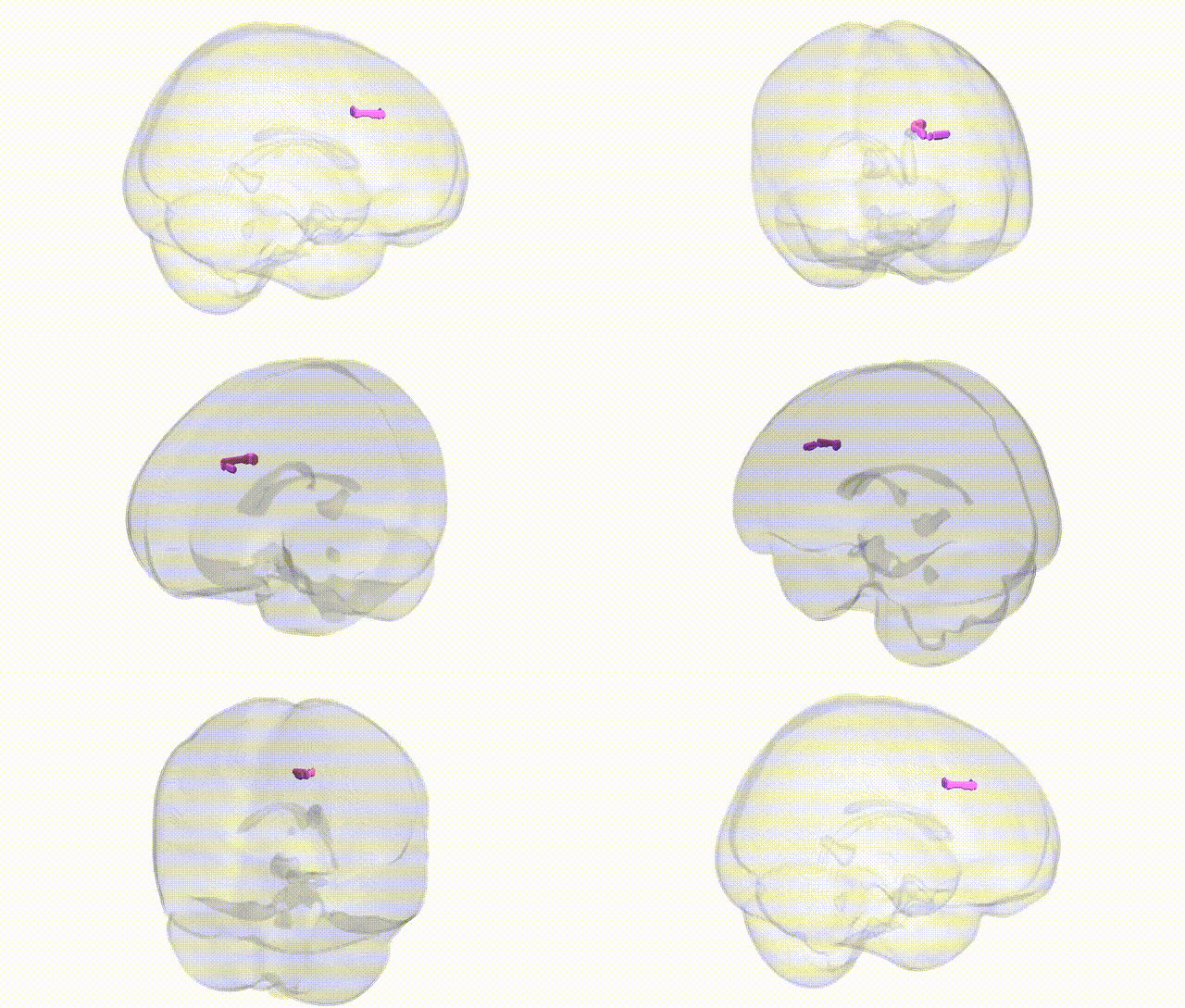
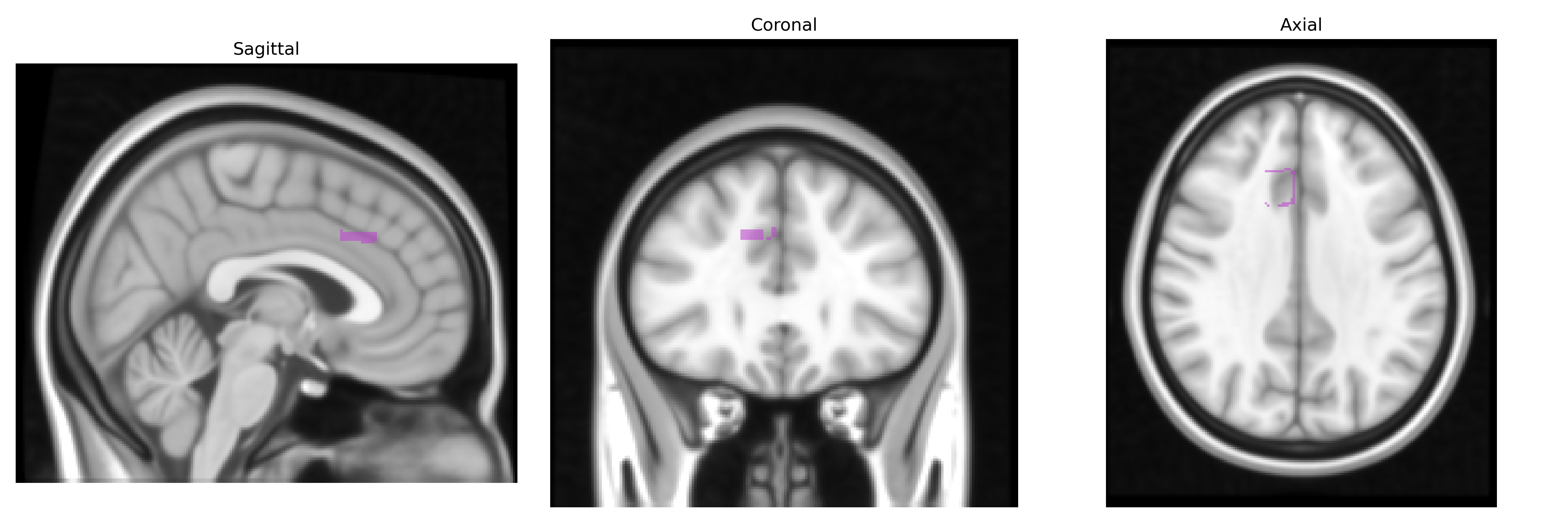
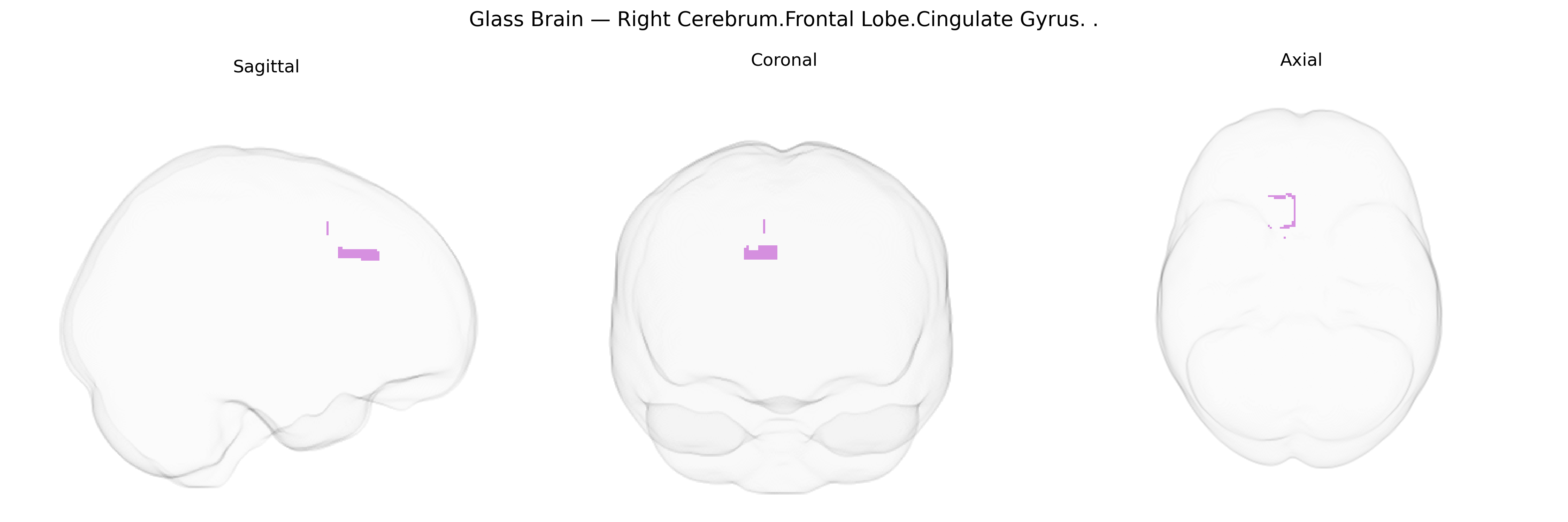
The bilateral Right Cerebrum.Frontal Lobe.Cingulate Gyrus in the Talairach 1 mm atlas refers to right-hemispheric portions of the cingulate gyrus within the frontal lobe, typically encompassing anterior and mid-cingulate sectors involved in higher-order cognitive and affective functions. This region forms part of the medial frontal cortex and the limbic system, arching above the corpus callosum and interfacing with prefrontal, motor, and limbic structures. Functionally, it contributes to executive control, error monitoring, conflict processing, motivation, and the regulation of emotion and autonomic responses, and is often implicated in attention, decision-making, pain processing, and mood-related disorders. There is no direct Wikipedia article for this exact Talairach label; a closely related structure is the Cingulate gyrus.
Genetic associations involving the bilateral right frontal lobe cingulate gyrus (as approximated by Talairach 1 mm atlas labels) converge on pathways related to cognitive control, emotion regulation, pain processing, and psychiatric vulnerability, with many findings centered on the anterior and mid-cingulate sectors. Large-scale GWAS and imaging‑genetics studies have linked common variants in genes such as COMT, DRD2, SLC6A4, BDNF, and DISC1, among others, to cingulate structure (e.g., cortical thickness, surface area, volume) and function (e.g., activation during conflict monitoring, error detection, and reward anticipation), often in a lateralized or regionally specific manner that encompasses the right frontal cingulate. Polygenic risk scores for schizophrenia, major depressive disorder, bipolar disorder, and ADHD have been associated with altered cingulate morphology or connectivity, while risk variants in the MHC region, CACNA1C, ZNF804A, and other psychiatric GWAS loci show intermediate phenotypes in cingulate-based networks. In neurodegenerative and neurodevelopmental conditions, APOE ε4, TREM2, and other Alzheimer’s disease risk variants correlate with cingulate hypometabolism or atrophy, and autism- and OCD‑related loci have been tied to atypical cingulate connectivity or activation patterns. Pain‑related GWAS (including variants near OPRM1 and genes involved in inflammatory and nociceptive signaling) implicate cingulate activity in chronic pain susceptibility, while individual differences in traits such as neuroticism, anxiety, impulsivity, and cognitive performance have been linked to cingulate measures in twin and SNP‑based heritability studies, indicating that genetic influences on this region contribute to a broad spectrum of behavioral and clinical phenotypes.
Overview generated by GPT-4o (2026).
Region ID: 1007
Hemisphere: bilateral
Atlas: Talairach labels 1mm
Full Quality Version: Download MP4
Full Quality Version: Download MP4
Wali Sidiqyar*, Gaurav Rudravaram*, Elyssa M. McMaster, Trent M. Schwartz, Adam M. Saunders, Kurt G. Schilling, Bennett A. Landman "Introducing SPINS: A Shared Public Visualization Library of Neuroanatomical Structures." Medical Imaging with Deep Learning- short paper
This resource is licensed under CC0 1.0 Universal (Public Domain).