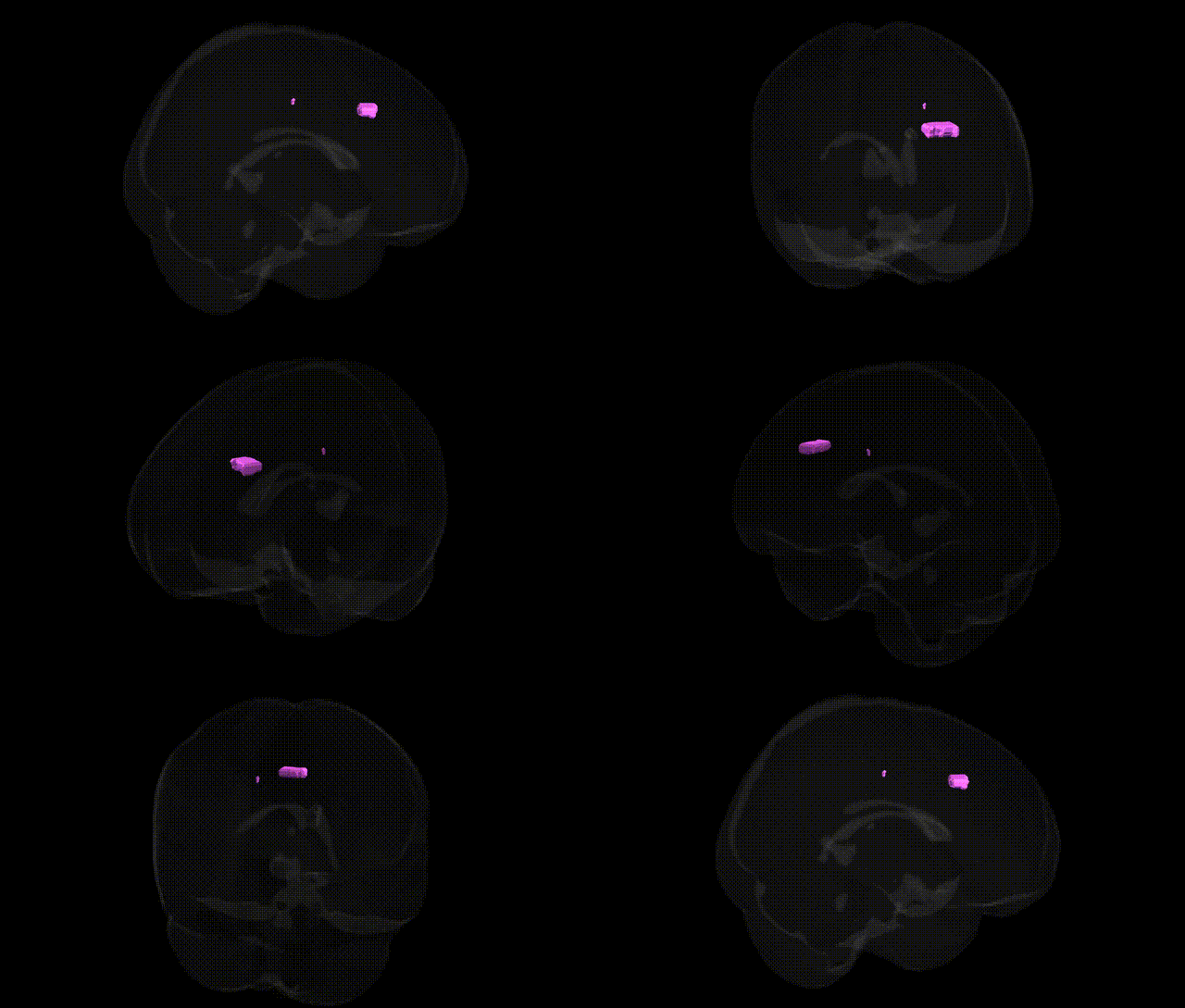
The bilateral Right Cerebrum.Frontal Lobe.Cingulate Gyrus.White Matter corresponds to subcortical myelinated fiber pathways running beneath the cingulate gyrus in the frontal portion of the cingulate cortex, which arches over the corpus callosum on the medial surface of the cerebral hemisphere. These white matter tracts connect anterior cingulate and medial prefrontal cortical regions with other limbic, frontal, and subcortical structures, supporting functions such as emotion regulation, motivation, decision-making, and cognitive control. As part of the broader cingulum bundle and frontal white matter, this region contributes to large-scale networks involved in attention, conflict monitoring, and the integration of affective and executive processes. There is no direct link for this exact Talairach-defined white matter label; a related structure is the Cingulate cortex.
Genetic associations involving the white matter of the cingulate gyrus in the right frontal lobe (and homologous left-sided regions) emerge primarily from imaging-genetics and GWAS of white matter microstructure, cingulum tract integrity, and cingulate volume rather than from Talairach-based studies directly; nonetheless, several robust links are relevant. Large diffusion MRI GWAS have identified multiple loci (for example near genes such as WNT3, DCC, and NTRK3) that influence fractional anisotropy and mean diffusivity in cingulum and adjacent frontal white matter, implicating axon guidance, myelination, and neurodevelopmental pathways. Variants in genes regulating myelin and oligodendrocyte function (e.g., MAG, MOG, and broader lipid metabolism loci) have been associated with global and regional white matter integrity, including cingulate-adjacent tracts, and overlap with genetic risk for multiple sclerosis and leukodystrophies. Polygenic risk for schizophrenia, major depressive disorder, bipolar disorder, and ADHD has been repeatedly linked to altered cingulate and frontal white matter microstructure and volume, with specific risk loci in synaptic and glutamatergic genes (such as CACNA1C, GRIN2A, and complement pathway genes) showing mediation effects on these structural measures. Cingulate white matter integrity is also influenced by Alzheimer’s disease and frontotemporal dementia risk loci (e.g., APOE, MAPT), which contribute to early white matter changes in limbic and frontal networks. In addition, GWAS of pain sensitivity, cognitive control, and neuroticism have found that polygenic scores for these traits predict cingulate/frontal white matter metrics, suggesting that distributed genetic architectures for affective and cognitive traits partly exert their effects through structural connectivity in the bilateral frontal cingulate white matter.
Overview generated by GPT-4o (2026).
Region ID: 1002
Hemisphere: bilateral
Atlas: Talairach labels 1mm
Full Quality Version: Download MP4
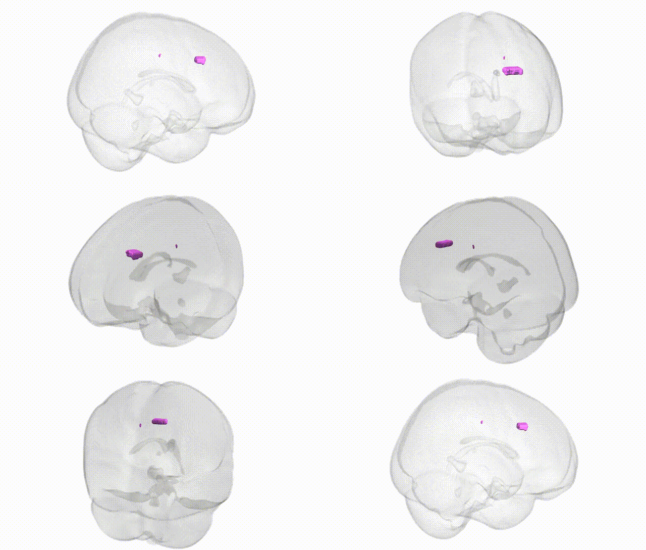
Full Quality Version: Download MP4
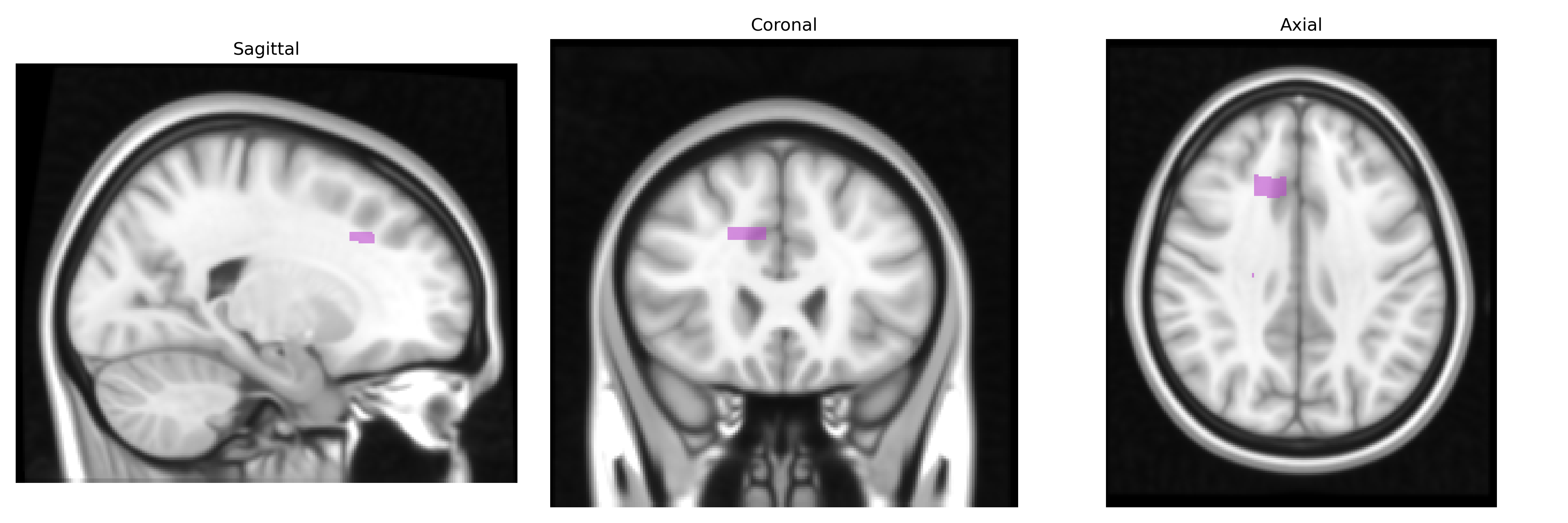
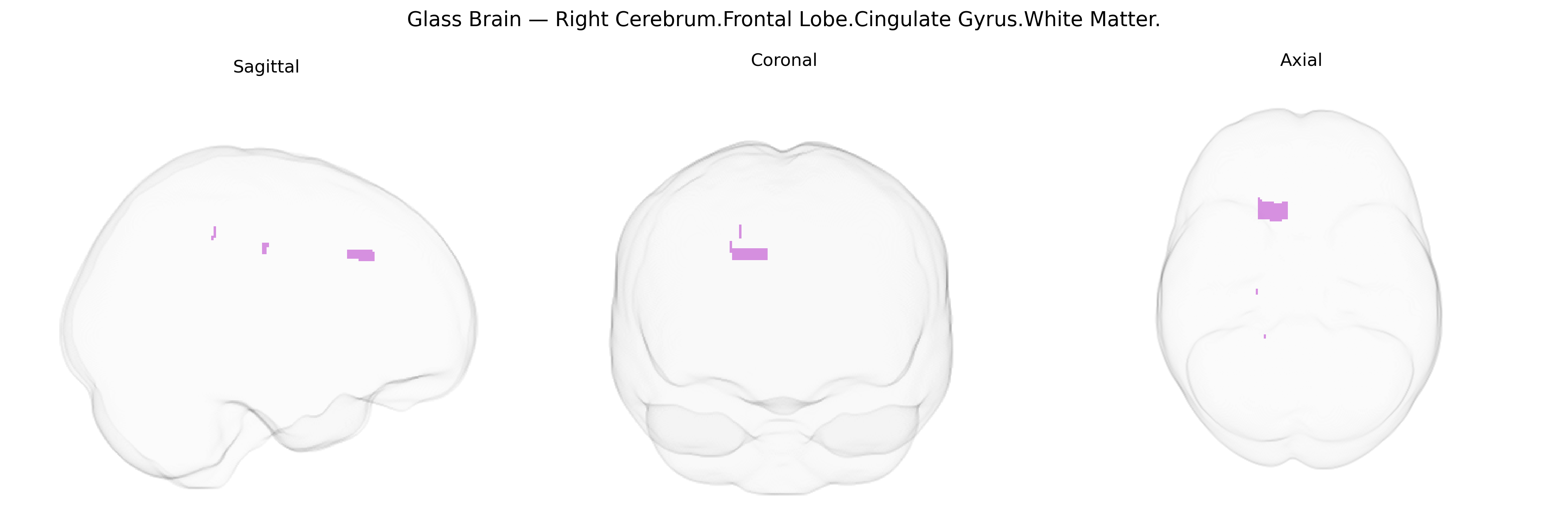
Wali Sidiqyar*, Gaurav Rudravaram*, Elyssa M. McMaster, Trent M. Schwartz, Adam M. Saunders, Kurt G. Schilling, Bennett A. Landman "Introducing SPINS: A Shared Public Visualization Library of Neuroanatomical Structures." Medical Imaging with Deep Learning- short paper
This resource is licensed under CC0 1.0 Universal (Public Domain).