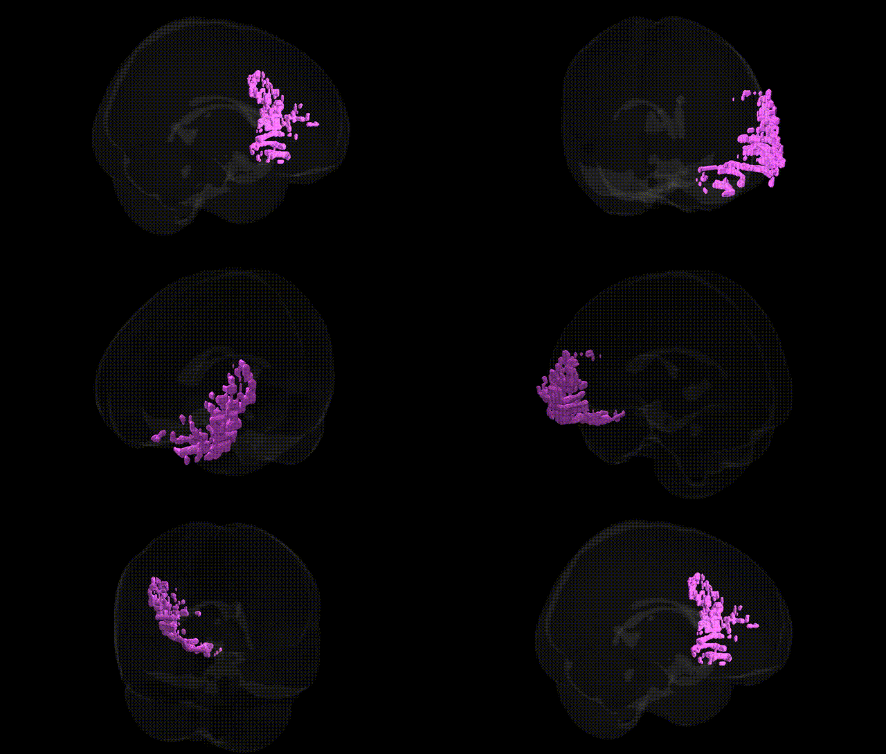
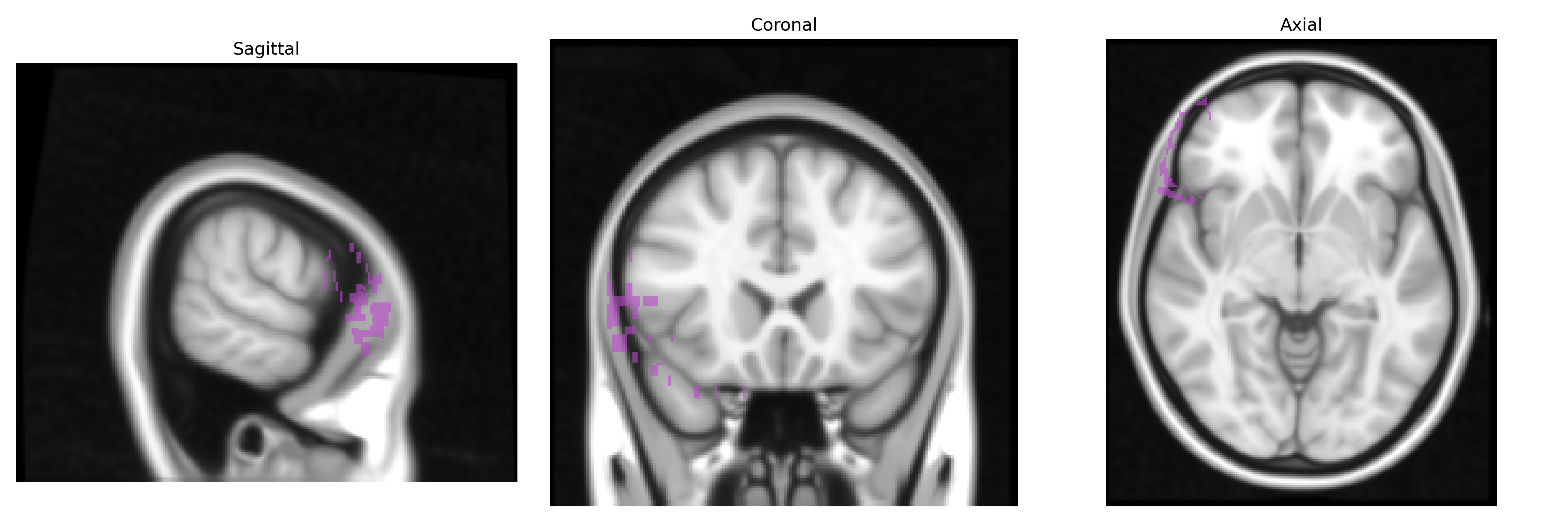
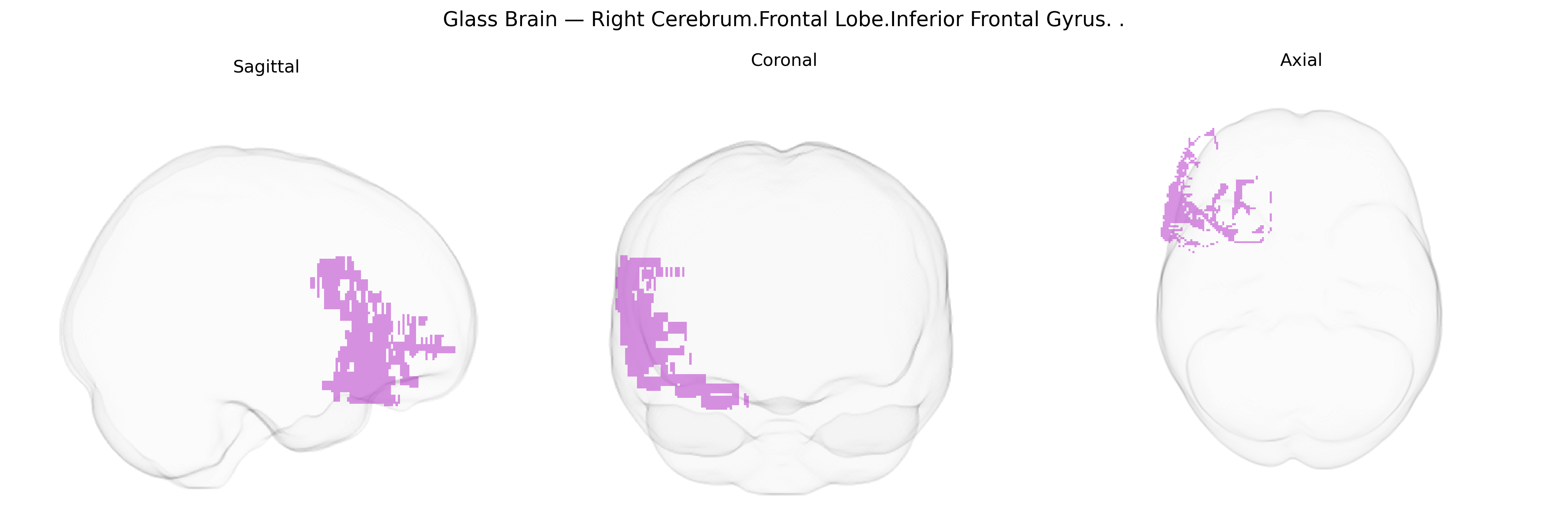
The bilateral Right Cerebrum.Frontal Lobe.Inferior Frontal Gyrus corresponds to the inferior frontal gyrus of the right frontal lobe, a cortical region implicated in higher-order cognitive and motor functions, including language, response inhibition, action observation, and aspects of social cognition. Anatomically, it is situated inferior to the middle frontal gyrus and above the lateral sulcus, and is often subdivided into pars opercularis, pars triangularis, and pars orbitalis based on cytoarchitectonic and functional criteria. Functionally, the right inferior frontal gyrus is strongly associated with inhibitory control of behavior (e.g., stopping or overriding prepotent responses), aspects of prosody and nonverbal components of communication, and participation in distributed networks for attention and executive function; it also overlaps with regions commonly described as part of the mirror neuron system. There is no Talairach-specific Wikipedia entry for this label, but a closely related article is: Inferior frontal gyrus.
The bilateral right inferior frontal gyrus (IFG) of the frontal lobe—corresponding to regions such as pars opercularis and pars triangularis in the Talairach 1 mm atlas—has been implicated in multiple genetic association studies, particularly those examining executive function, response inhibition, language, and psychiatric or neurodevelopmental disorders. Imaging–genetics work and GWAS of cortical morphology have linked common variants in genes involved in synaptic plasticity and neurodevelopment (for example, BDNF, COMT, NRG1, and CNTNAP2) to structural and functional differences in IFG, including gray matter volume and activation patterns during tasks requiring inhibitory control and language processing. Large-scale cortical GWAS consortia (e.g., ENIGMA, UK Biobank–based studies) have identified loci affecting frontal cortical thickness and surface area that map to genes regulating neuronal migration, axon guidance, and myelination, some of which show strongest or significant effects in inferior frontal regions and are shared with risk loci for schizophrenia, bipolar disorder, major depression, and ADHD. Right IFG activation and structure have been repeatedly associated with variants influencing dopamine and serotonin signaling (including DRD2/ANKK1 and SLC6A4), which modulate impulsivity, response inhibition, and risk-taking traits. Additionally, genetic risk for autism spectrum disorder, dyslexia, and language impairment has been associated with altered IFG morphology or connectivity, and polygenic scores for cognitive ability and educational attainment show correlations with IFG measures in population neuroimaging cohorts, supporting a role for this region as a structural and functional mediator of genetically influenced differences in higher-order cognition and self-control.
Overview generated by GPT-4o (2026).
Region ID: 187
Hemisphere: bilateral
Atlas: Talairach labels 1mm
Full Quality Version: Download MP4
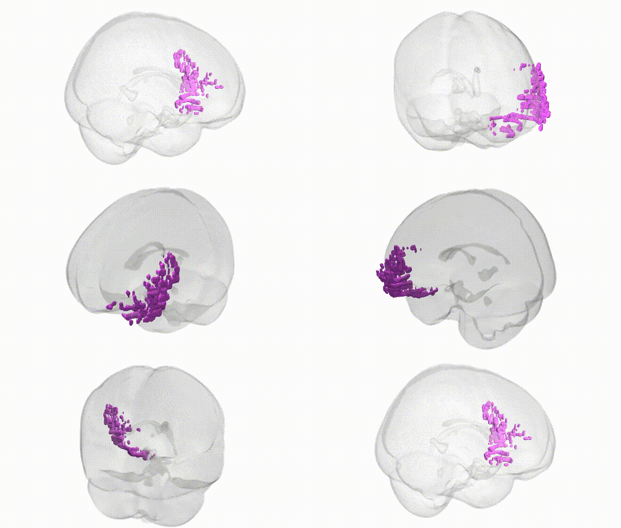
Full Quality Version: Download MP4
Wali Sidiqyar*, Gaurav Rudravaram*, Elyssa M. McMaster, Trent M. Schwartz, Adam M. Saunders, Kurt G. Schilling, Bennett A. Landman "Introducing SPINS: A Shared Public Visualization Library of Neuroanatomical Structures." Medical Imaging with Deep Learning- short paper
This resource is licensed under CC0 1.0 Universal (Public Domain).