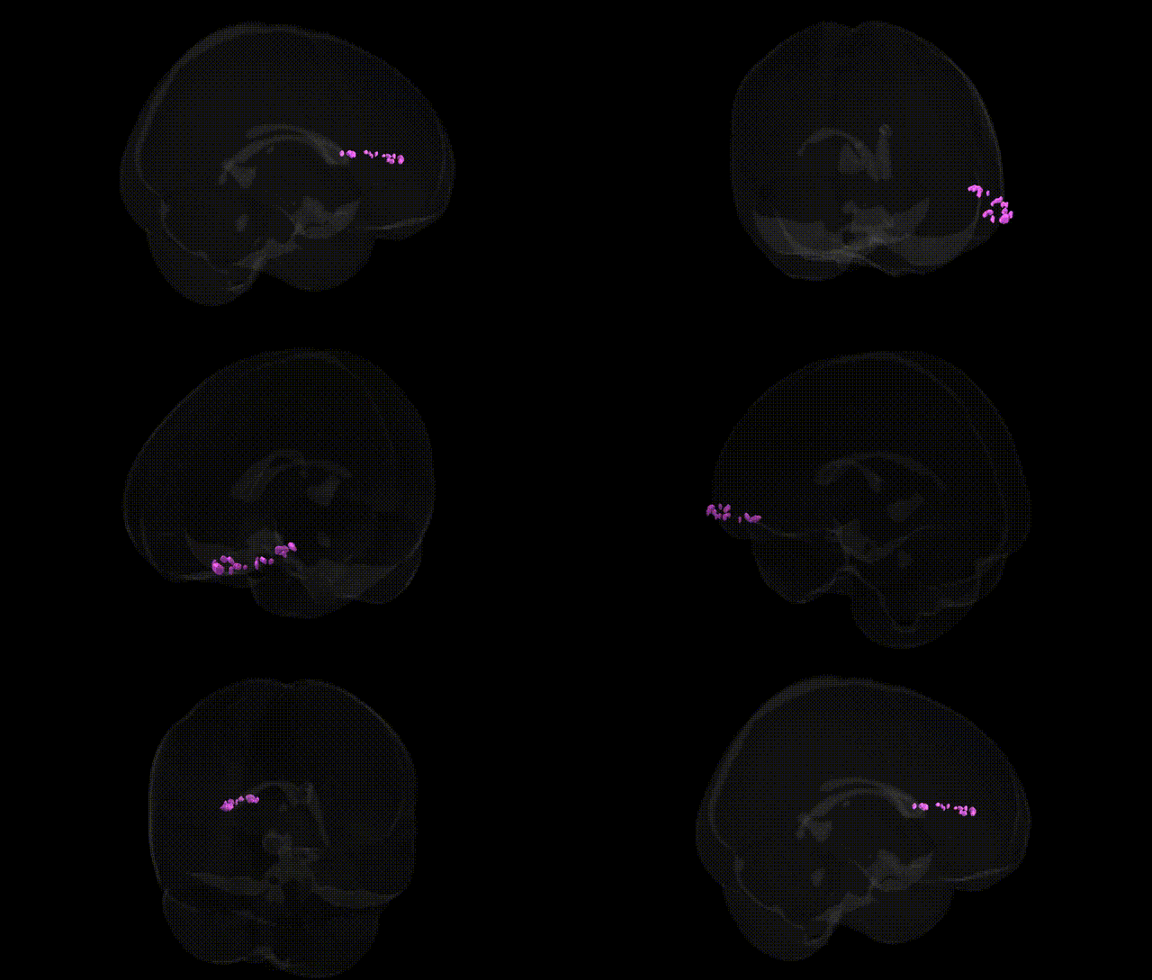
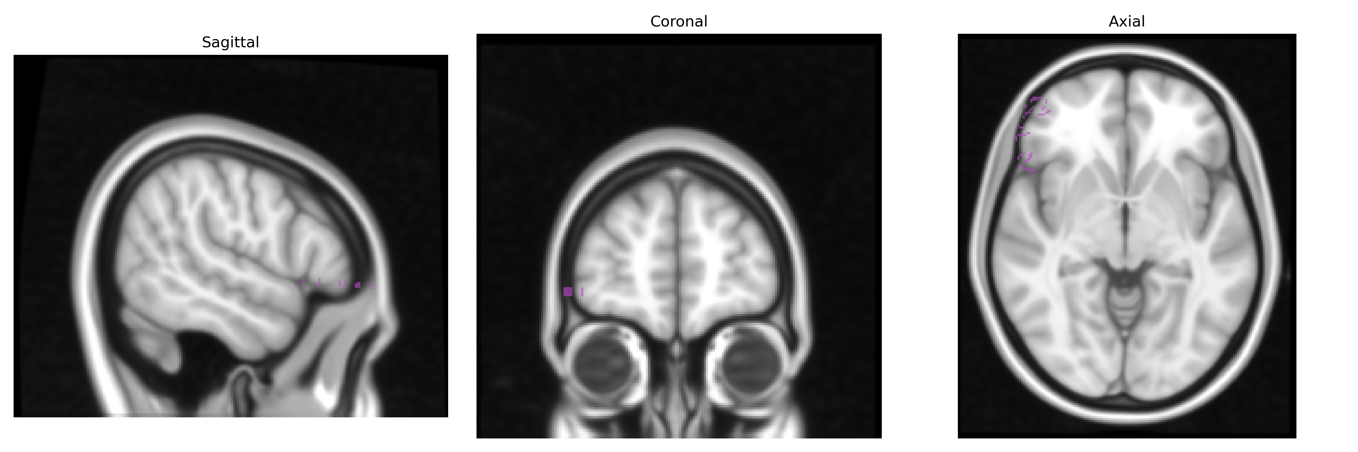
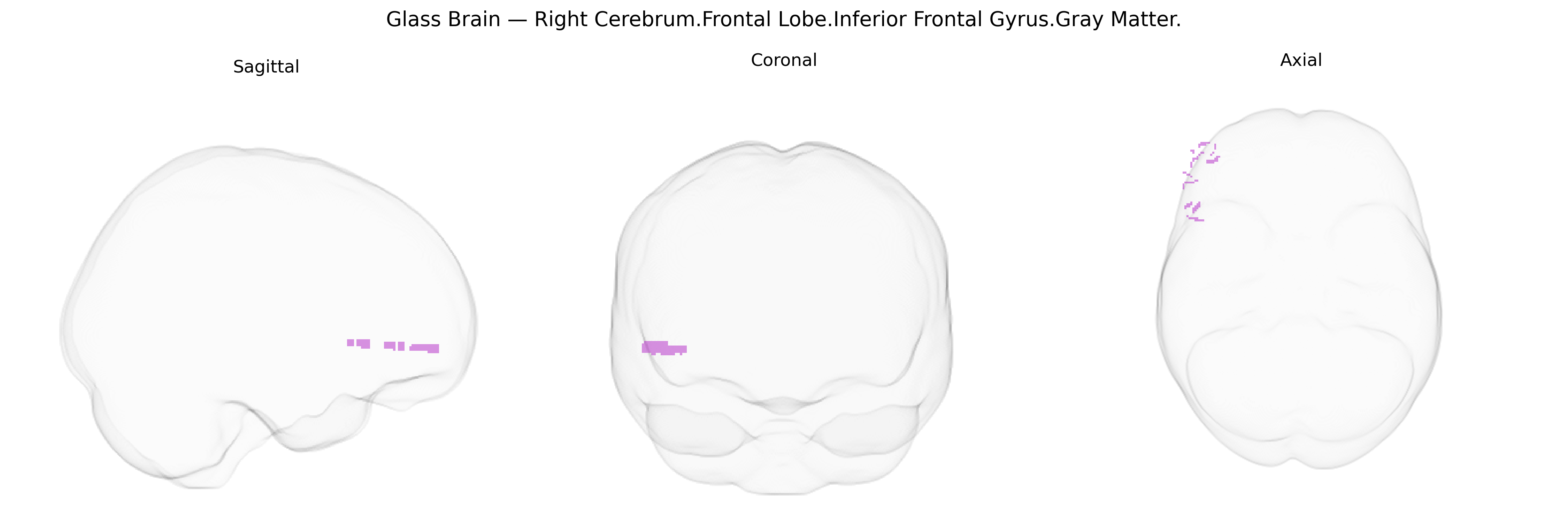
The bilateral Right Cerebrum.Frontal Lobe.Inferior Frontal Gyrus.Gray Matter corresponds to cortical tissue in the inferior frontal gyrus of the right frontal lobe, a region implicated in higher-order cognitive and language-related processes, motor planning, and aspects of social cognition. This area includes parts of the pars opercularis, pars triangularis, and pars orbitalis, which participate in networks for response inhibition, working memory, and the modulation of speech and prosody, often complementing left-hemisphere language functions. Functionally, the right inferior frontal gyrus has been associated with attention control, suppression of inappropriate actions, and interpretation of emotional tone in language (prosody), integrating sensory, motor, and affective information to support flexible, context-dependent behavior.
The bilateral right inferior frontal gyrus (IFG) gray matter, as defined in the Talairach 1 mm atlas, has been implicated in multiple genetic and GWAS-based associations primarily through imaging genetics and large-scale neuroimaging consortia such as ENIGMA and UK Biobank. Variants in genes influencing synaptic development and cortical structure (e.g., BDNF, CNTNAP2, FOXP2, and common polygenic influences captured by cortical thickness/volume GWAS) have been linked to inter-individual differences in IFG morphology and activation, particularly in tasks involving response inhibition, language, and social cognition. GWAS of cortical surface area and thickness have identified numerous loci (including regions near KIAA0586, C15orf53, and others) associated with frontal lobe metrics that encompass or overlap the IFG. Clinically, genetic risk for attention-deficit/hyperactivity disorder, obsessive–compulsive disorder, and substance use disorders has been associated with altered structure or function of the right IFG, often in the context of inhibitory control networks. Polygenic scores for schizophrenia, bipolar disorder, and major depressive disorder show correlations with frontal cortical morphometry and connectivity patterns that include the inferior frontal regions, and autism-related variants and language-related genes (such as FOXP2) have been connected to atypical development of IFG-associated circuits, though most findings are distributed and not specific to this single Talairach-defined parcel. Overall, genetic associations with this region are largely polygenic and network-based, with right IFG anatomy and function serving as an intermediate phenotype linking common and rare variants to cognitive control, language, and neuropsychiatric vulnerability.
Overview generated by GPT-4o (2026).
Region ID: 600
Hemisphere: bilateral
Atlas: Talairach labels 1mm
Full Quality Version: Download MP4
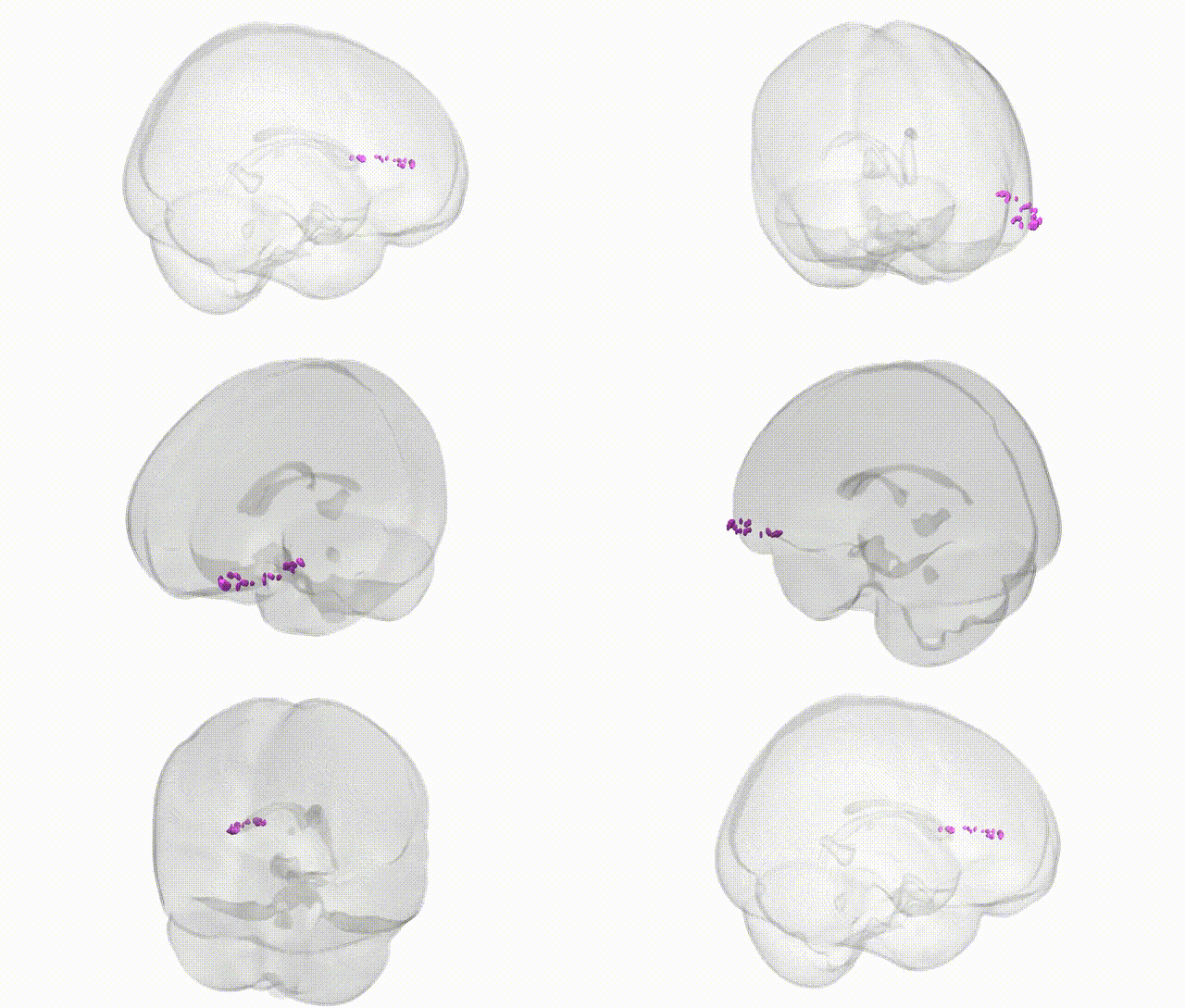
Full Quality Version: Download MP4
Wali Sidiqyar*, Gaurav Rudravaram*, Elyssa M. McMaster, Trent M. Schwartz, Adam M. Saunders, Kurt G. Schilling, Bennett A. Landman "Introducing SPINS: A Shared Public Visualization Library of Neuroanatomical Structures." Medical Imaging with Deep Learning- short paper
This resource is licensed under CC0 1.0 Universal (Public Domain).