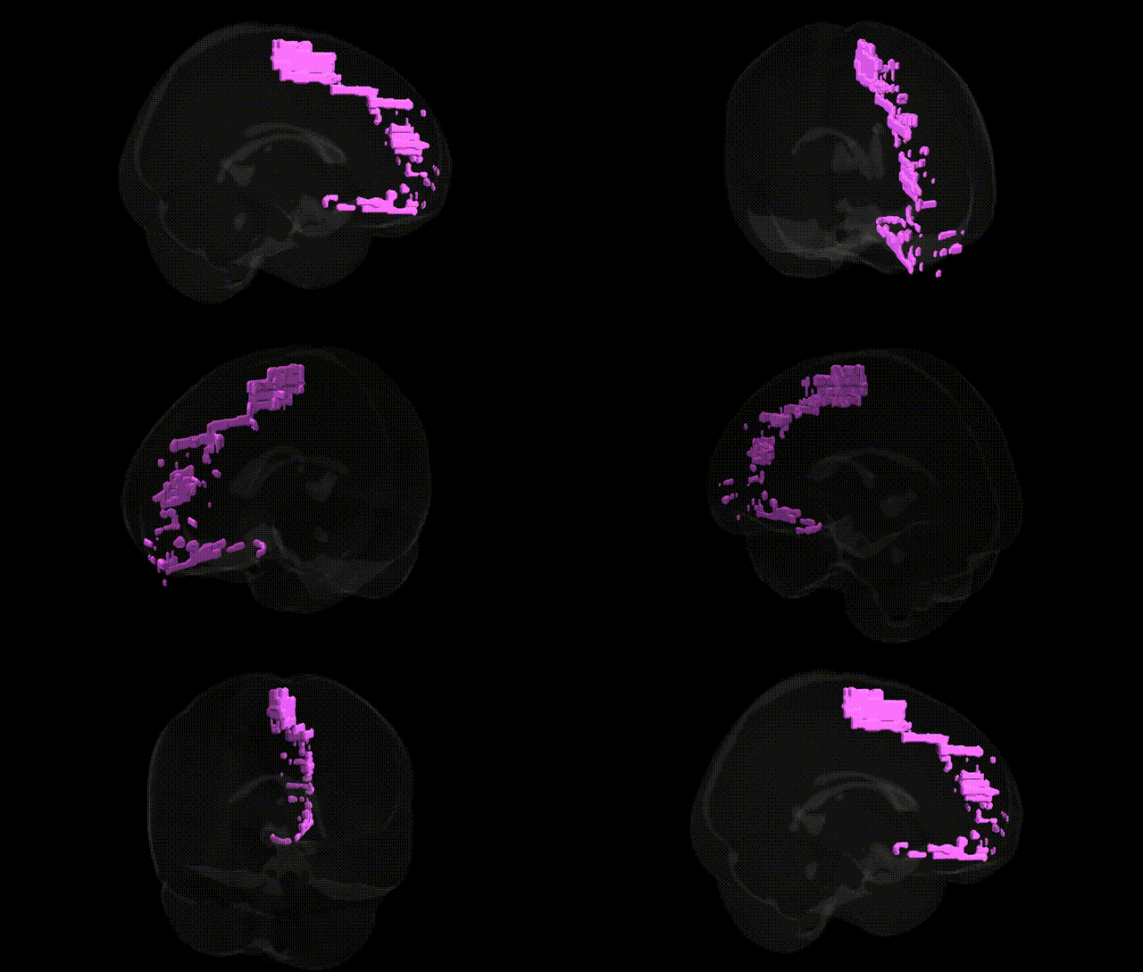
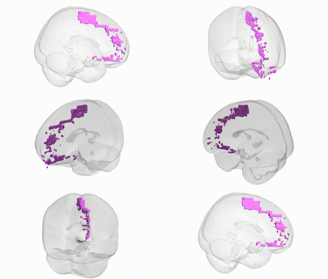
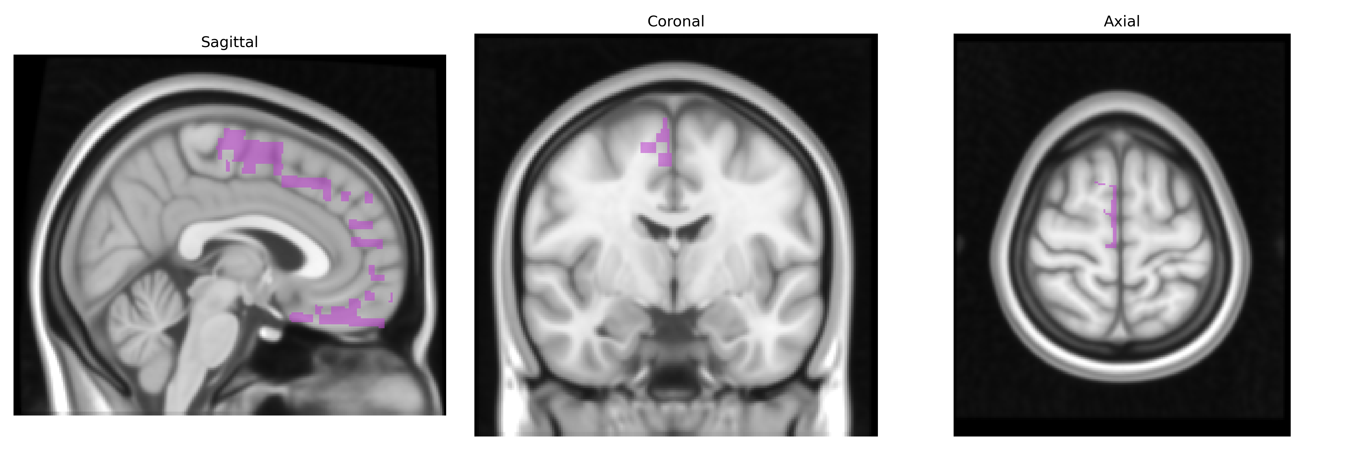
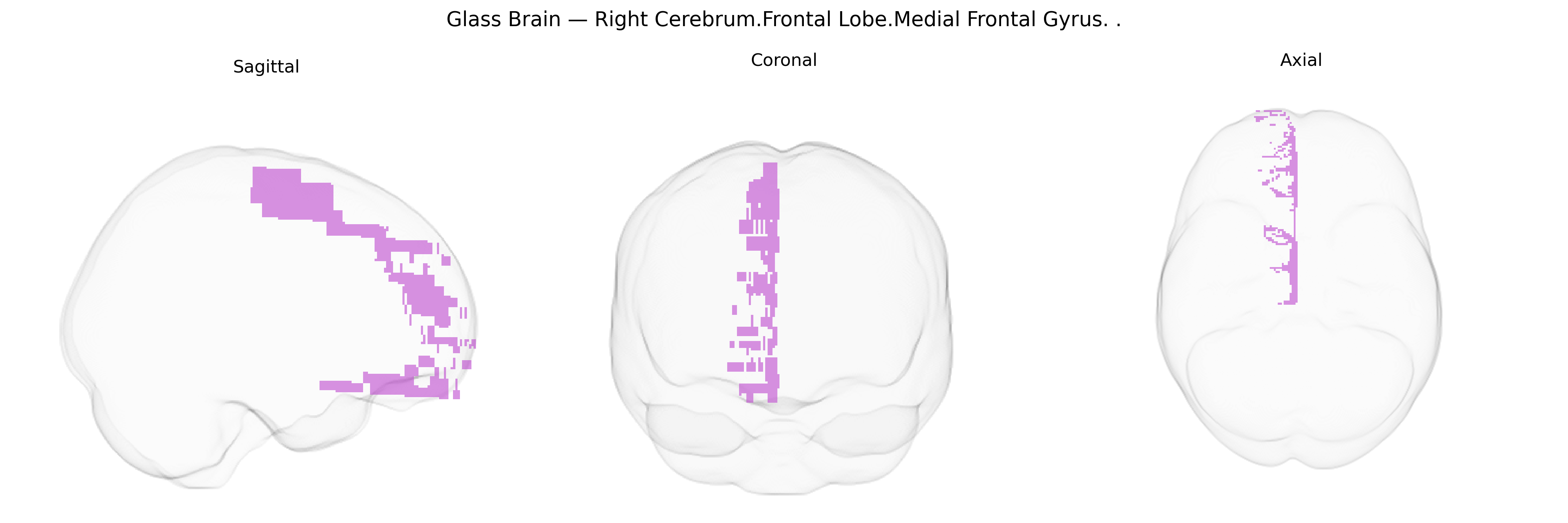
The bilateral Right Cerebrum.Frontal Lobe.Medial Frontal Gyrus corresponds primarily to the medial portion of the superior frontal gyrus along the mesial surface of the frontal lobe, extending anteriorly from the supplementary motor area toward the frontal pole and bounded inferiorly by the cingulate sulcus. This region encompasses parts of Brodmann areas 8, 9, 10, and 32, with functional involvement in higher-order executive processes, attention, decision-making, and aspects of self-referential and internally directed cognition, as well as contributions to motor planning and performance monitoring via its connections with premotor, cingulate, and prefrontal networks. There is no direct link for “medial frontal gyrus”; see the related structure Superior frontal gyrus.
The medial frontal gyrus (bilateral right cerebrum frontal lobe, Talairach 1 mm atlas) has been implicated in multiple genetic and genome-wide association studies that link its structure and function to cognition, affective regulation, and psychiatric and neurologic disorders. GWAS of cortical thickness and surface area (e.g., ENIGMA and UK Biobank consortia) have identified common variants in genes related to neurodevelopment (such as MIR137, HMGA2, and others in synaptic and neuronal growth pathways) that influence medial prefrontal morphology and connectivity. Imaging–genetics work has associated medial frontal volume, thickness, and activation patterns with polygenic risk scores for schizophrenia, bipolar disorder, major depressive disorder, attention-deficit/hyperactivity disorder, and autism, including effects tied to risk loci in CACNA1C, ZNF804A, and DRD2, which modulate fronto-striatal and default-mode network function. Variants in serotonin transporter (SLC6A4) and COMT genes have been linked to altered medial frontal activation during emotion and cognitive control tasks, while APOE and other Alzheimer’s disease risk loci show associations with medial frontal atrophy and metabolic changes in aging cohorts. GWAS of behavioral and personality traits (such as educational attainment, intelligence, neuroticism, and risk-taking) also converge on medial prefrontal circuitry, with polygenic profiles influencing structural and functional metrics in this region, suggesting a shared genetic architecture spanning higher-order cognition, affect, and vulnerability to psychiatric illness.
Overview generated by GPT-4o (2026).
Region ID: 224
Hemisphere: bilateral
Atlas: Talairach labels 1mm
Full Quality Version: Download MP4
Full Quality Version: Download MP4
Wali Sidiqyar*, Gaurav Rudravaram*, Elyssa M. McMaster, Trent M. Schwartz, Adam M. Saunders, Kurt G. Schilling, Bennett A. Landman "Introducing SPINS: A Shared Public Visualization Library of Neuroanatomical Structures." Medical Imaging with Deep Learning- short paper
This resource is licensed under CC0 1.0 Universal (Public Domain).