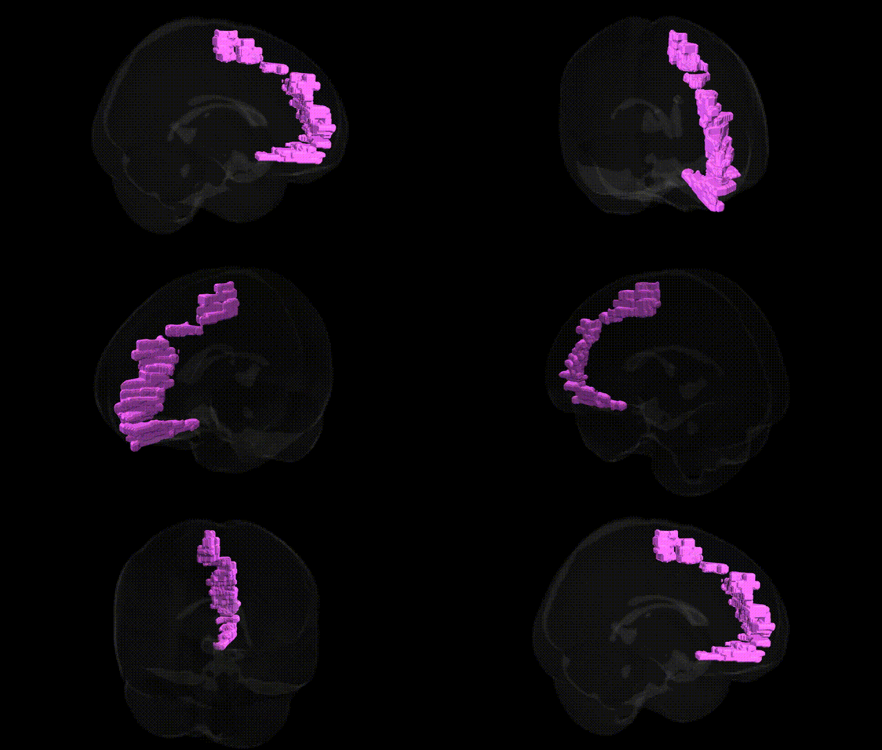
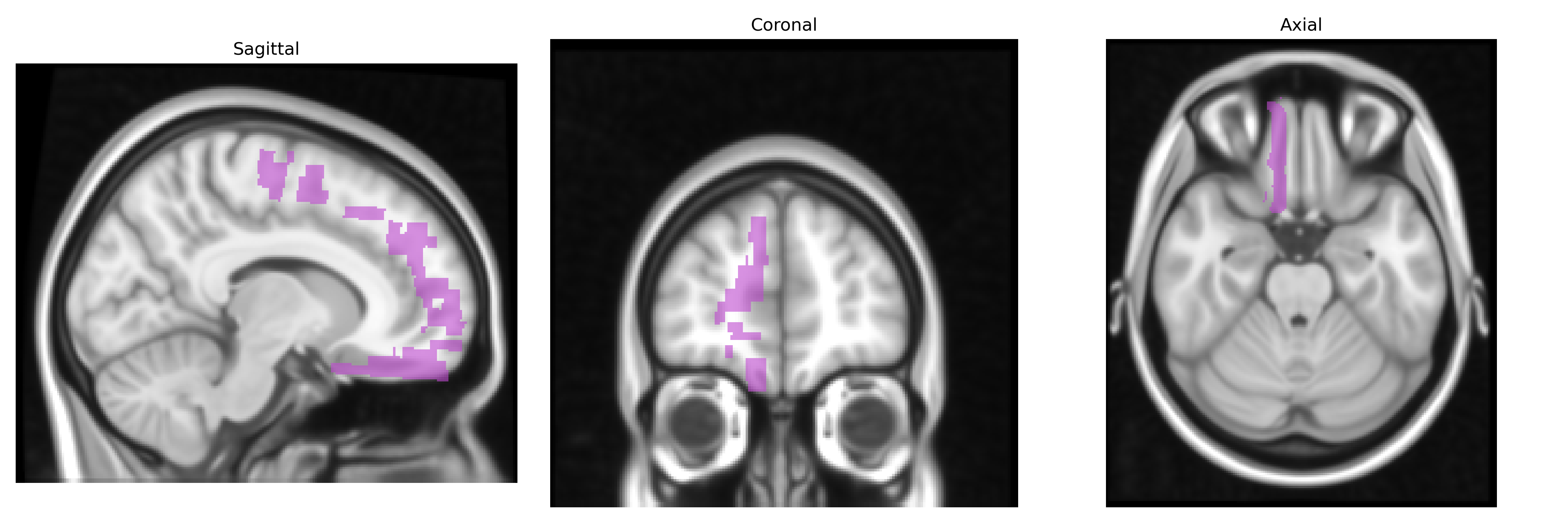
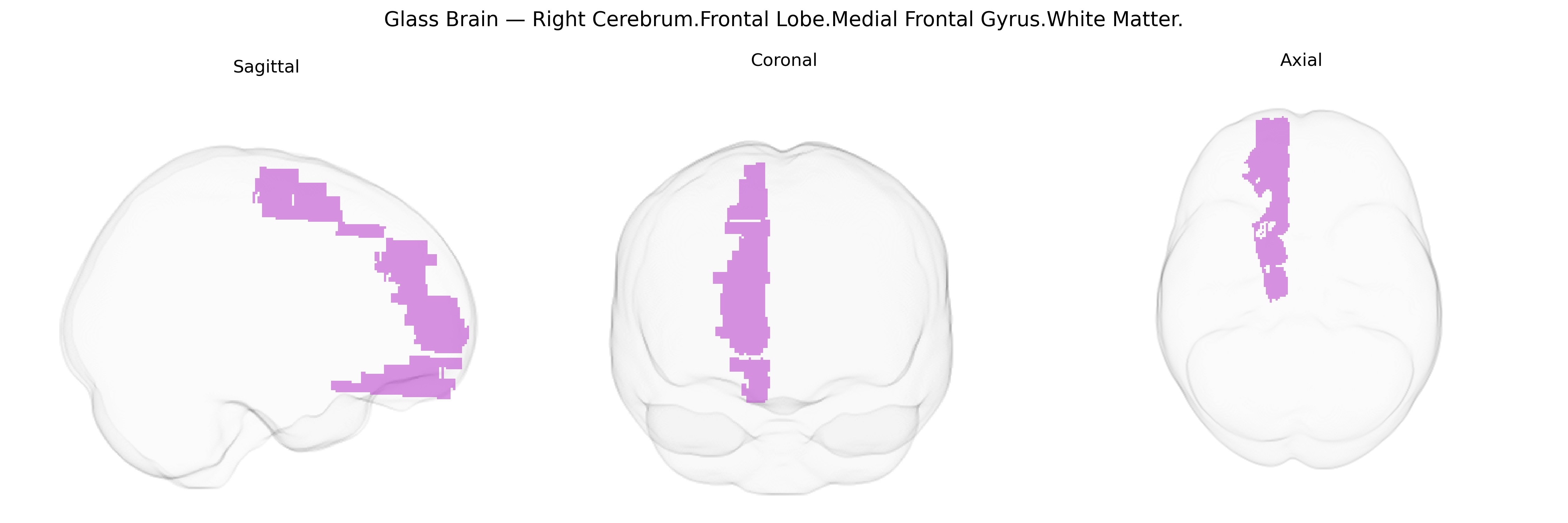
The bilateral Right Cerebrum.Frontal Lobe.Medial Frontal Gyrus.White Matter corresponds to the subcortical fiber pathways underlying the medial frontal gyrus, a portion of the medial prefrontal cortex situated on the medial surface of the frontal lobe. This white matter region contains association fibers connecting medial frontal cortex with other prefrontal areas, limbic structures (such as the cingulate cortex), and subcortical nuclei, supporting functions related to executive control, motivation, decision-making, and aspects of social and emotional behavior. As part of the broader frontal white matter network, it contributes to the integration of internally driven actions, performance monitoring, and regulation of goal-directed behavior. There is no direct Wikipedia article for this specific white matter subdivision; a closely related cortical region is the Medial frontal gyrus.
The bilateral right medial frontal gyrus white matter, as defined in the Talairach 1 mm atlas, corresponds broadly to medial prefrontal and anterior cingulate–adjacent tracts that have been implicated in multiple genetic imaging and GWAS studies of brain structure and function. Variants in genes influencing myelination, axonal growth, and synaptic signaling—such as NRG1, ERBB4, CNTNAP2, DISC1, and CACNA1C—have been associated with white matter integrity and connectivity in medial frontal regions, often via diffusion tensor imaging measures (e.g., fractional anisotropy) that overlap with this tract. Large-scale neuroimaging GWAS consortia (e.g., ENIGMA, UK Biobank) have identified polygenic influences on cortical thickness, surface area, and white matter microstructure in frontal midline regions, linking them to traits such as general cognitive ability, executive function, educational attainment, and personality dimensions like neuroticism and conscientiousness. Medial frontal white matter has also been repeatedly implicated in psychiatric and neurological disorders with known genetic components: schizophrenia and bipolar disorder (through risk loci including CACNA1C, ZNF804A, and multiple genome-wide significant variants), major depressive disorder, obsessive–compulsive disorder, ADHD, and autism spectrum disorder, with case–control and endophenotype studies reporting genotype-dependent differences in medial frontal connectivity. Additionally, genetic risk scores for Alzheimer’s disease, frontotemporal dementia, and vascular risk factors (e.g., APOE variants) show associations with frontal white matter integrity, suggesting that the right medial frontal gyrus white matter participates in genetically influenced networks supporting higher-order cognition, emotion regulation, and vulnerability to psychiatric and neurodegenerative conditions, even though many findings map to slightly broader medial prefrontal or anterior cingulate–centered regions rather than this Talairach label alone.
Overview generated by GPT-4o (2026).
Region ID: 231
Hemisphere: bilateral
Atlas: Talairach labels 1mm
Full Quality Version: Download MP4
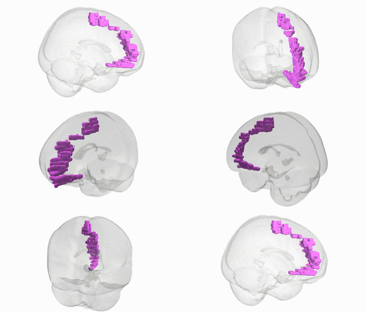
Full Quality Version: Download MP4
Wali Sidiqyar*, Gaurav Rudravaram*, Elyssa M. McMaster, Trent M. Schwartz, Adam M. Saunders, Kurt G. Schilling, Bennett A. Landman "Introducing SPINS: A Shared Public Visualization Library of Neuroanatomical Structures." Medical Imaging with Deep Learning- short paper
This resource is licensed under CC0 1.0 Universal (Public Domain).