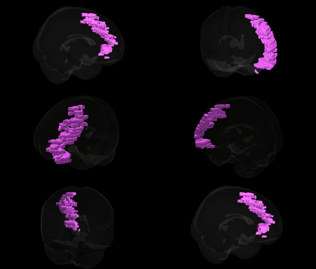
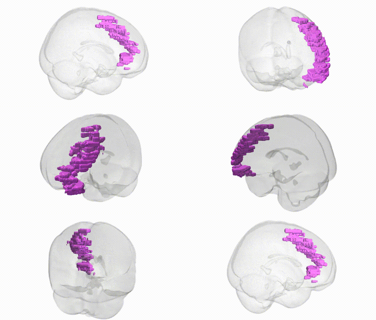
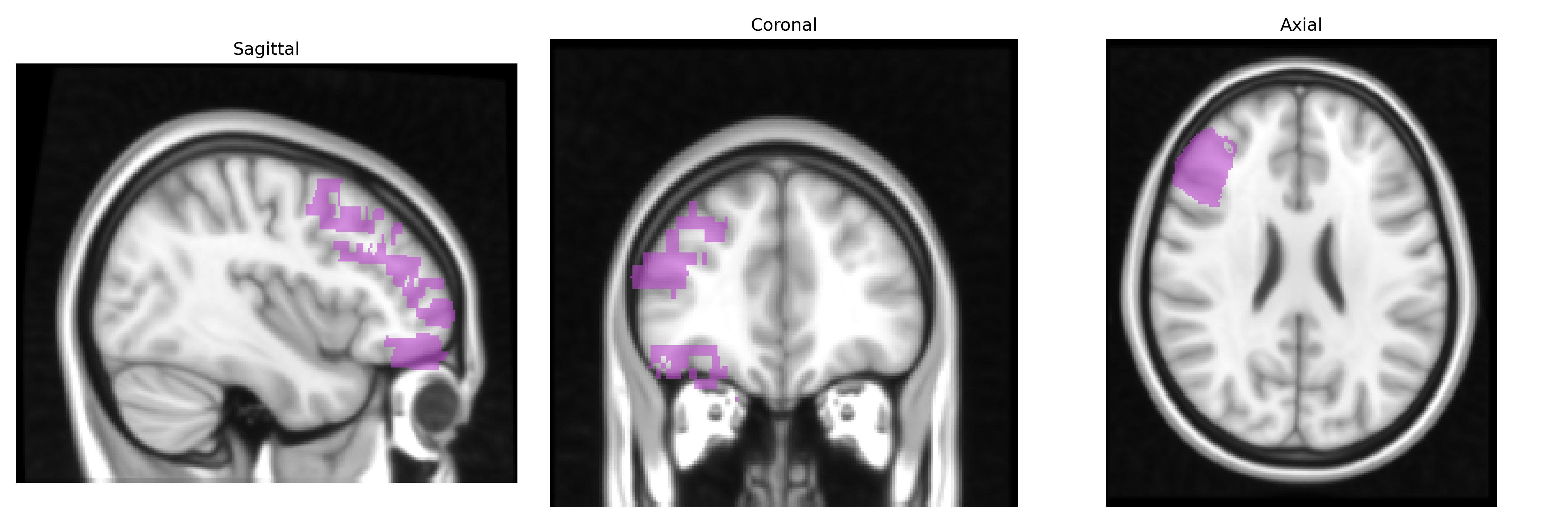
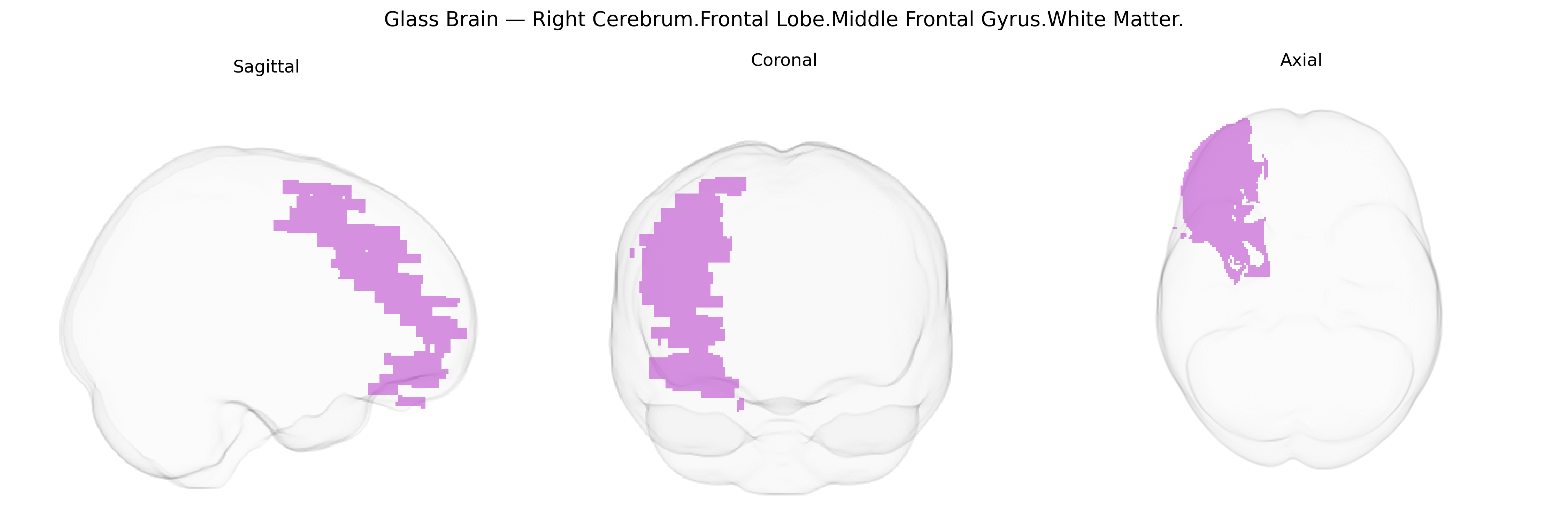
The bilateral Right Cerebrum.Frontal Lobe.Middle Frontal Gyrus.White Matter corresponds to the deep medullary white matter underlying the middle frontal gyrus in the right frontal lobe, containing association fibers that interconnect prefrontal cortical areas both within the frontal lobe and with distant cortical regions such as parietal and temporal association cortices. This white matter supports higher-order executive functions, including working memory, attentional control, planning, and decision-making, by facilitating efficient communication within the dorsolateral prefrontal network and between hemispheres via callosal fibers. It also carries descending fibers that influence premotor and motor regions, thereby linking cognitive control processes to action selection and motor planning. There is no direct Wikipedia article for this specific white matter subdivision; a related structure is the Middle Frontal Gyrus.
Genetic associations involving the bilateral Right Cerebrum.Frontal Lobe.Middle Frontal Gyrus.White Matter, as defined in the Talairach 1 mm atlas, largely arise from imaging genetics and GWAS of structural and diffusion MRI measures of frontal white matter integrity and volume rather than from studies targeting this exact label. Common variants in genes affecting myelination, axon guidance, and synaptic plasticity—such as those in CNTNAP2, NRG1, DISC1, and CACNA1C—have been repeatedly implicated in frontal lobe white matter microstructure and connectivity, which includes tracts coursing through the middle frontal gyrus region. GWAS of global and regional frontal white matter volume and fractional anisotropy (FA) have identified loci near genes like LINGO1, BDNF, and multiple oligodendrocyte-related genes, with effects overlapping association and executive networks anchored in the middle frontal gyrus. These genetic influences on frontal white matter have been linked to neuropsychiatric disorders including schizophrenia, bipolar disorder, major depressive disorder, ADHD, and autism spectrum disorder, as well as cognitive traits such as working memory, intelligence, and educational attainment, through intermediate phenotypes such as altered dorsolateral prefrontal connectivity and fronto-striatal circuitry. While no large GWAS has focused specifically on the Talairach-defined “Right Middle Frontal Gyrus White Matter” label, convergent evidence indicates that variants influencing frontal lobe development and myelination affect white matter architecture in this region and thereby contribute to risk or resilience for disorders characterized by executive dysfunction and altered prefrontal network organization.
Overview generated by GPT-4o (2026).
Region ID: 196
Hemisphere: bilateral
Atlas: Talairach labels 1mm
Full Quality Version: Download MP4
Full Quality Version: Download MP4
Wali Sidiqyar*, Gaurav Rudravaram*, Elyssa M. McMaster, Trent M. Schwartz, Adam M. Saunders, Kurt G. Schilling, Bennett A. Landman "Introducing SPINS: A Shared Public Visualization Library of Neuroanatomical Structures." Medical Imaging with Deep Learning- short paper
This resource is licensed under CC0 1.0 Universal (Public Domain).