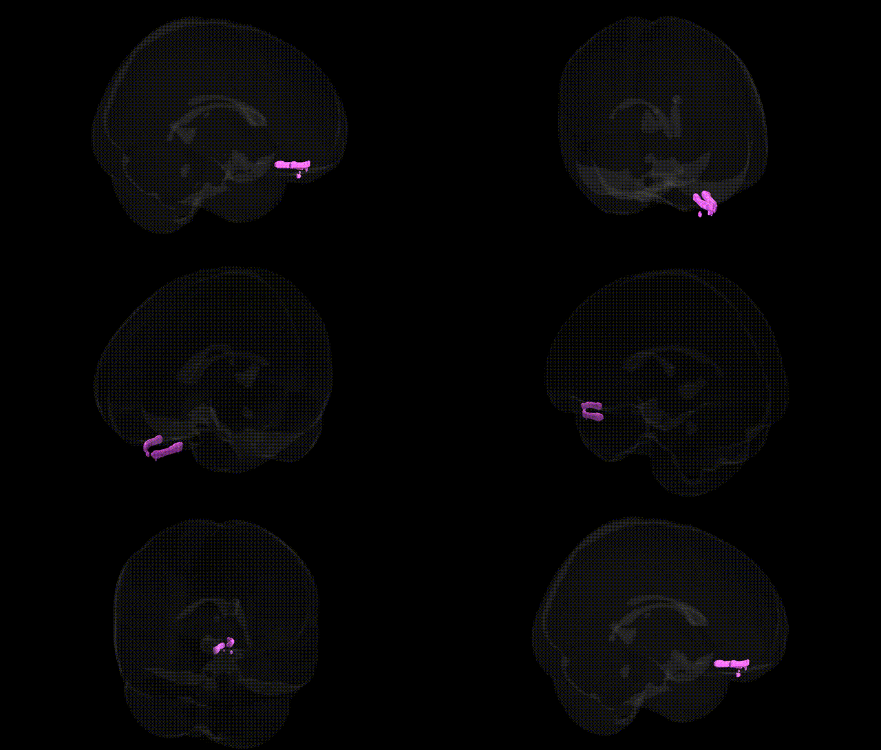
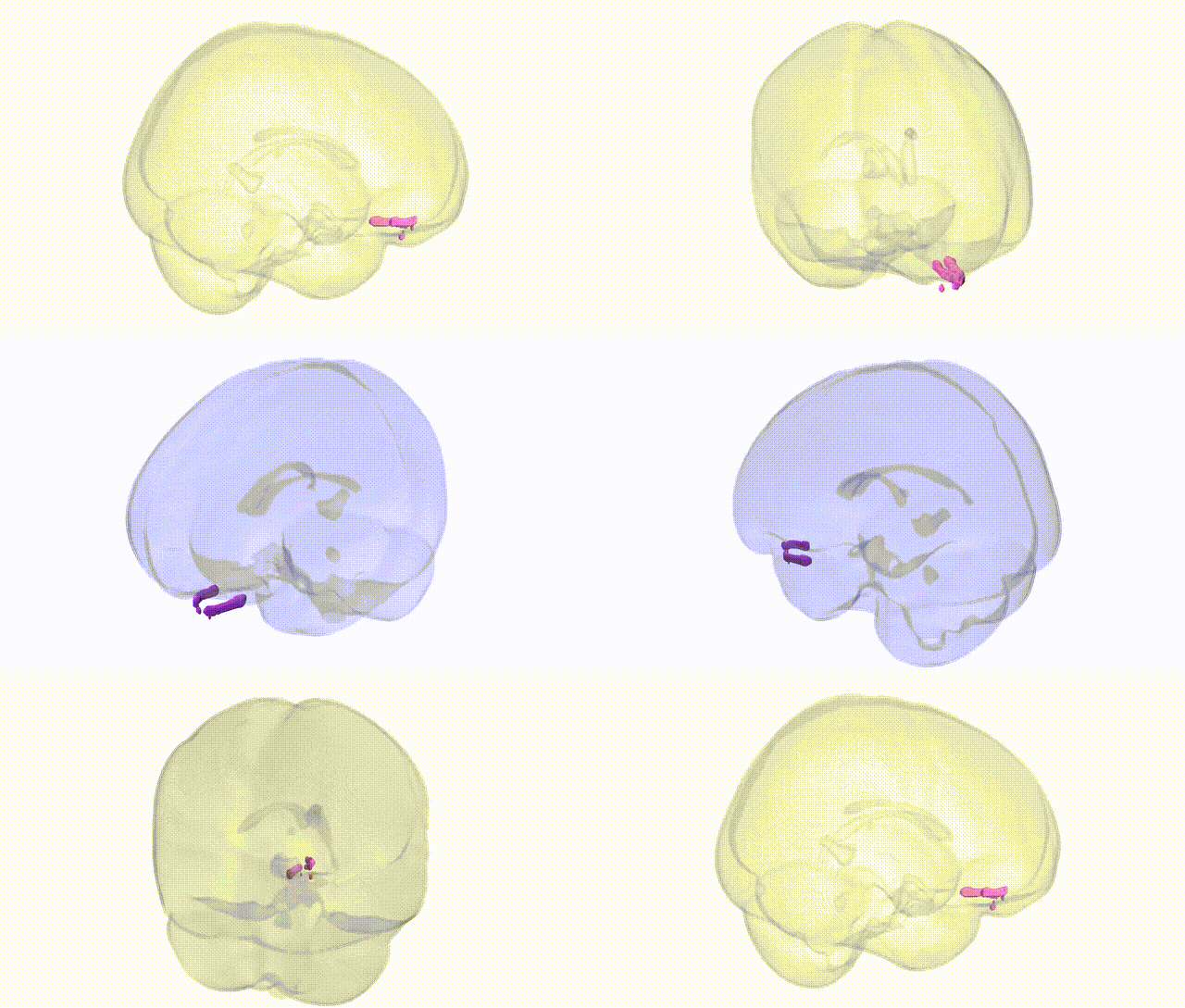
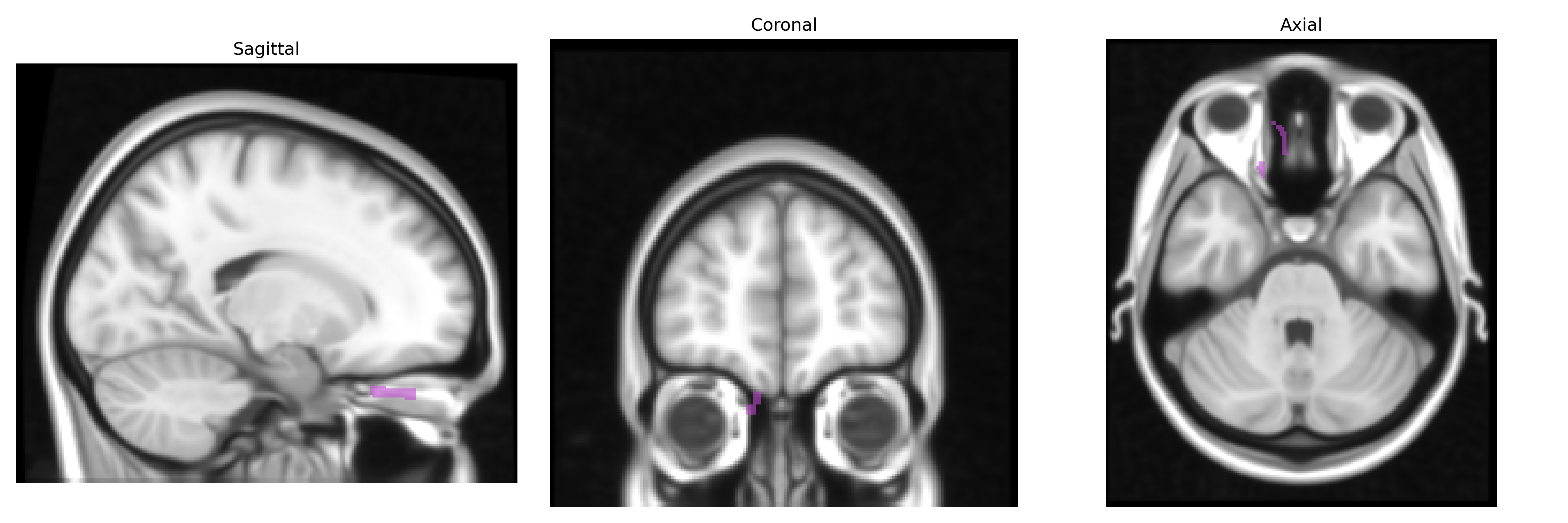
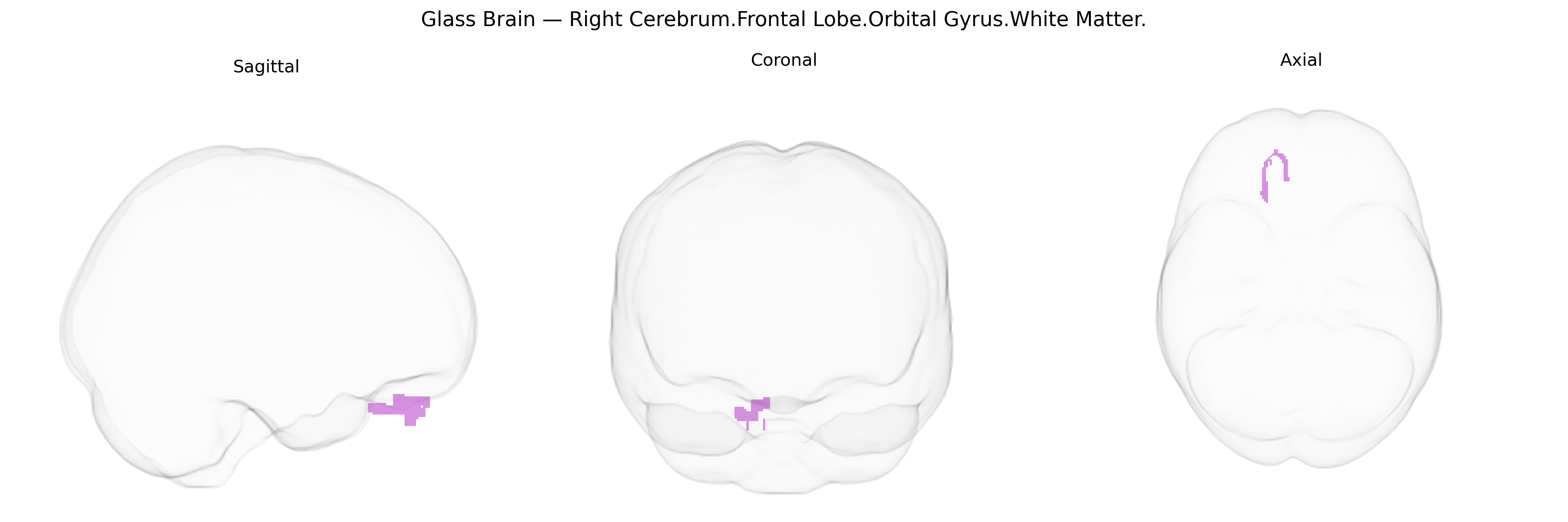
The bilateral Right Cerebrum.Frontal Lobe.Orbital Gyrus.White Matter corresponds to the subcortical fiber systems underlying the orbital gyri of the frontal lobe on the right side, including interconnecting association fibers that link orbitofrontal cortex with other prefrontal, limbic, and subcortical regions. These white matter pathways help support functions attributed to the overlying orbitofrontal cortex, such as reward evaluation, decision-making, emotional regulation, and aspects of social behavior, by transmitting processed sensory, affective, and contextual information. As part of the inferior frontal lobe’s deep structure, this region participates in networks involving the limbic system (including amygdala and ventral striatum) and other prefrontal territories, contributing to the integration of motivational and cognitive signals. There is no direct link for this exact white matter label; a related cortical structure is the Orbitofrontal Cortex.
The bilateral right orbital frontal white matter (Right Cerebrum.Frontal Lobe.Orbital Gyrus.White Matter in the Talairach 1 mm atlas) is indirectly implicated in genetic associations primarily through imaging genetics and psychiatric GWAS that map risk variants to structural and connectivity alterations in orbitofrontal circuitry rather than to this exact white-matter parcel per se. Large-scale GWAS of brain structure (e.g., ENIGMA and UK Biobank imaging cohorts) have identified common variants in genes related to neurodevelopment, axon guidance, and myelination—such as NRG1/NRG3, NCAM1, ROBO/SLIT pathways, and oligodendrocyte-related genes—associated with global and regional frontal lobe white-matter integrity (often measured by fractional anisotropy), with effects that include the ventral/orbitofrontal sector. Psychiatric and behavioral GWAS for major depression, bipolar disorder, schizophrenia, obsessive-compulsive disorder, substance use, and neuroticism frequently highlight polygenic burdens that converge on fronto-limbic and orbitofrontal networks, and imaging–genetic mediation analyses show that risk loci in genes such as CACNA1C, DRD2, and GRM3 relate to altered orbitofrontal volume or connectivity, including adjacent white-matter tracts. Additionally, variants in genes regulating serotonin and dopamine signaling (e.g., SLC6A4, COMT) and lipid/myelin biology (e.g., PLP1, MOG) have been associated with differences in frontal white-matter microstructure and impulsivity or decision-making phenotypes typically linked to orbitofrontal function. However, current evidence is regionally coarse, and no major GWAS has yet isolated robust, highly specific associations uniquely and reproducibly confined to the bilateral right orbital frontal white matter as defined by this particular Talairach label; instead, genetic effects are distributed across broader orbitofrontal and fronto-subcortical white-matter systems.
Overview generated by GPT-4o (2026).
Region ID: 113
Hemisphere: bilateral
Atlas: Talairach labels 1mm
Full Quality Version: Download MP4
Full Quality Version: Download MP4
Wali Sidiqyar*, Gaurav Rudravaram*, Elyssa M. McMaster, Trent M. Schwartz, Adam M. Saunders, Kurt G. Schilling, Bennett A. Landman "Introducing SPINS: A Shared Public Visualization Library of Neuroanatomical Structures." Medical Imaging with Deep Learning- short paper
This resource is licensed under CC0 1.0 Universal (Public Domain).