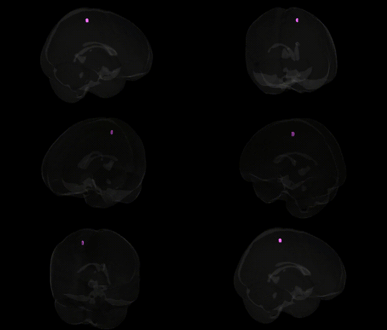
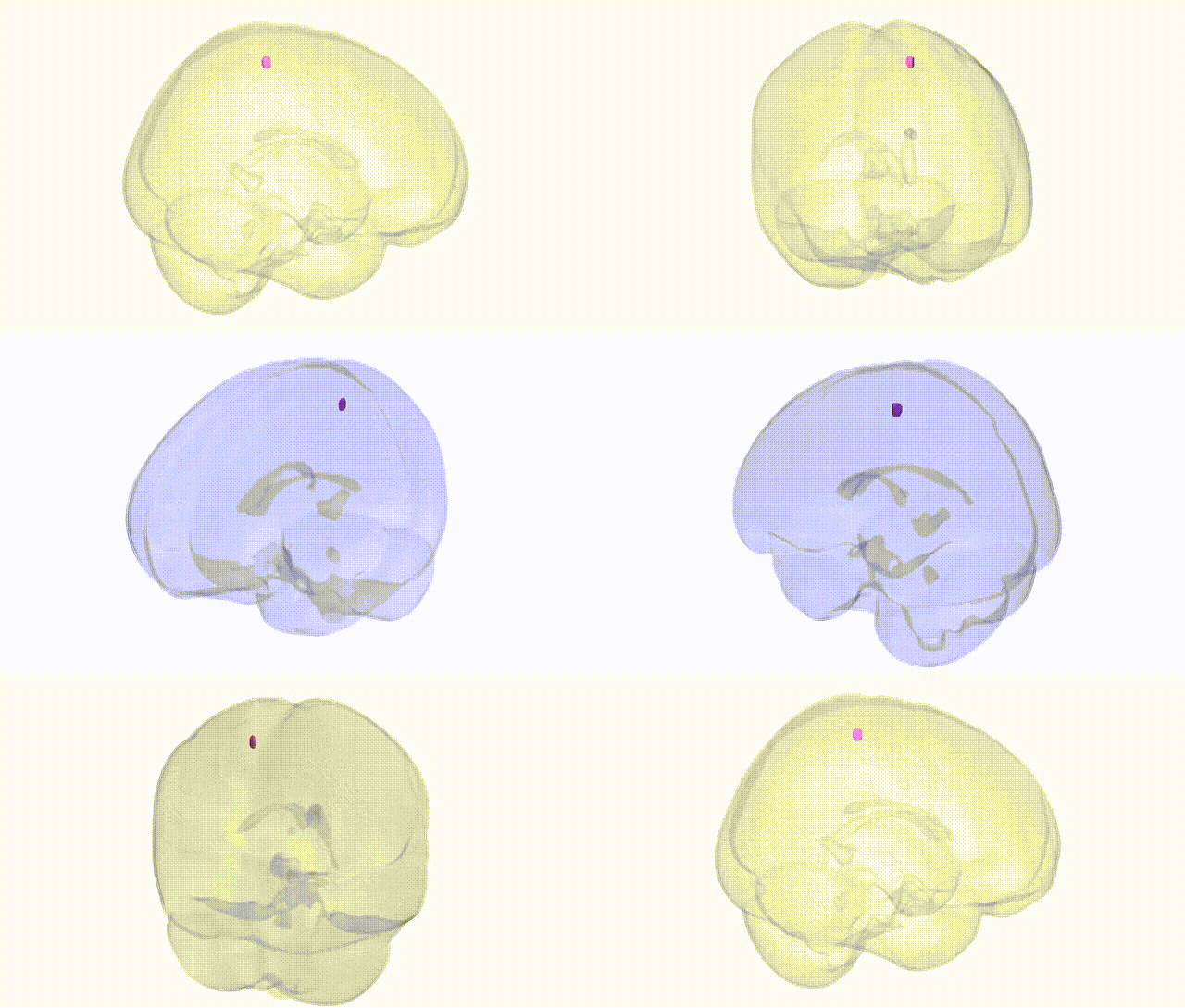
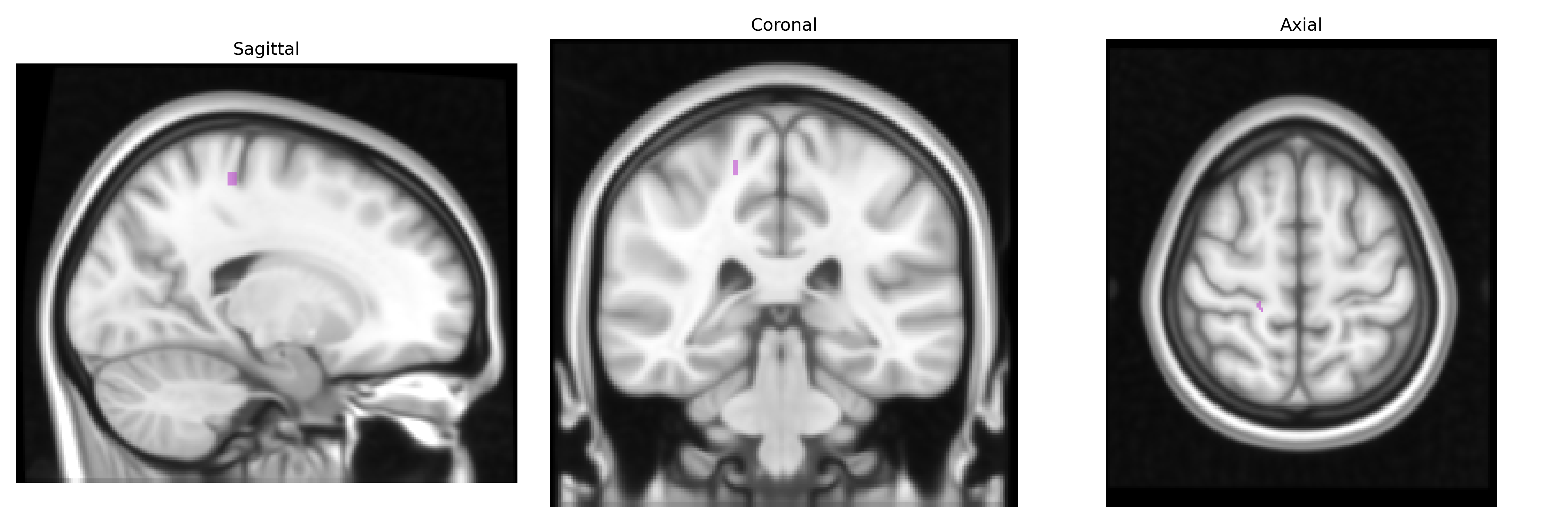
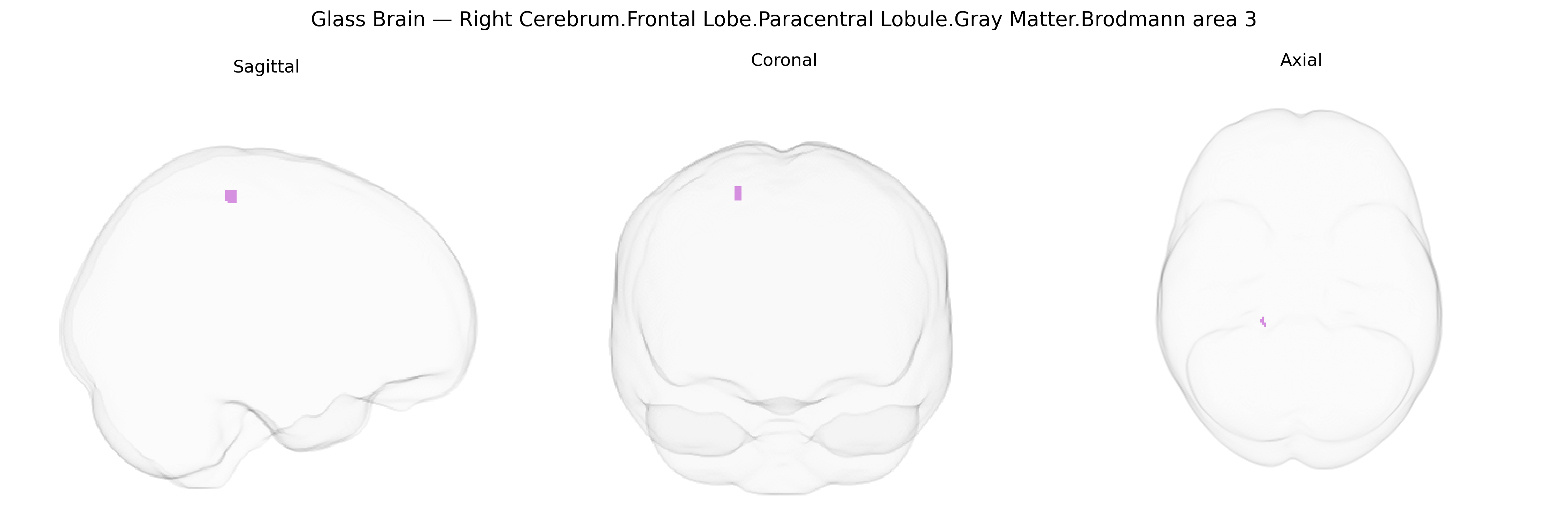
The bilateral Right Cerebrum.Frontal Lobe.Paracentral Lobule.Gray Matter.Brodmann area 3 corresponds to primary somatosensory cortex (S1) territory located along the posterior portion of the paracentral lobule on the medial surface of the frontal lobe, immediately anterior to the central sulcus and extending into the interhemispheric fissure. This region is cytoarchitectonically defined by Brodmann as area 3, which is heavily granular and characterized by dense thalamocortical input from the ventral posterior nuclei, and is primarily involved in processing fine tactile, proprioceptive, and nociceptive information from the contralateral body, especially from the lower limb and perineal regions represented medially in the somatotopic “homunculus.” Functionally, it serves as an initial cortical recipient of somatosensory signals that are further relayed to adjacent Brodmann areas 1 and 2 for higher-order integration, contributing to conscious perception of touch, position sense, and body schema. There is no direct link for this exact composite label; a closely related structure is the primary somatosensory cortex: Primary somatosensory cortex.
The bilateral right paracentral lobule gray matter in Brodmann area 3, a primary somatosensory subregion in the medial frontal lobe, has been implicated in several genetic and GWAS-based associations through its roles in sensorimotor processing, pain, and motor control. Large-scale imaging–genetics consortia (e.g., ENIGMA, UK Biobank) have identified common variants influencing cortical thickness, surface area, and gyrification of the paracentral lobule, with prominent loci including variants near genes involved in neurodevelopment and synaptic signaling (such as MIR137, HMGA2, ZIC4, and genes in Wnt and axon-guidance pathways), although these are usually reported at the regional or sensorimotor cortex level rather than isolated to right BA3. GWAS of chronic pain and somatosensory traits have associated variants in genes related to nociceptive processing (e.g., TRPV1, CACNA1H, SCN9A) with altered activation or structural variation in primary somatosensory areas that encompass BA3, while motor and gait-related GWAS (including Parkinson’s disease, amyotrophic lateral sclerosis, and polygenic risk for motor impairment) show downstream effects on paracentral lobule function via corticospinal and sensorimotor circuits. Structural and functional MRI studies stratified by genotype link polygenic risk for multiple sclerosis, epilepsy, and autism spectrum conditions to altered morphology or connectivity of medial sensorimotor cortex, including the paracentral lobule, and schizophrenia risk variants (e.g., in complement pathway genes like C4A and synaptic genes such as GRIN2A) have been associated with widespread cortical thickness changes that include primary sensory regions. Overall, genetic associations involving this region are typically captured at the level of primary sensorimotor or medial frontal cortex rather than a unique, region-specific signature for right cerebrum BA3, with most evidence pointing to polygenic influences on its structure and function across neurodevelopmental, psychiatric, and movement- or pain-related traits.
Overview generated by GPT-4o (2026).
Region ID: 1091
Hemisphere: bilateral
Atlas: Talairach labels 1mm
Full Quality Version: Download MP4
Full Quality Version: Download MP4
Wali Sidiqyar*, Gaurav Rudravaram*, Elyssa M. McMaster, Trent M. Schwartz, Adam M. Saunders, Kurt G. Schilling, Bennett A. Landman "Introducing SPINS: A Shared Public Visualization Library of Neuroanatomical Structures." Medical Imaging with Deep Learning- short paper
This resource is licensed under CC0 1.0 Universal (Public Domain).