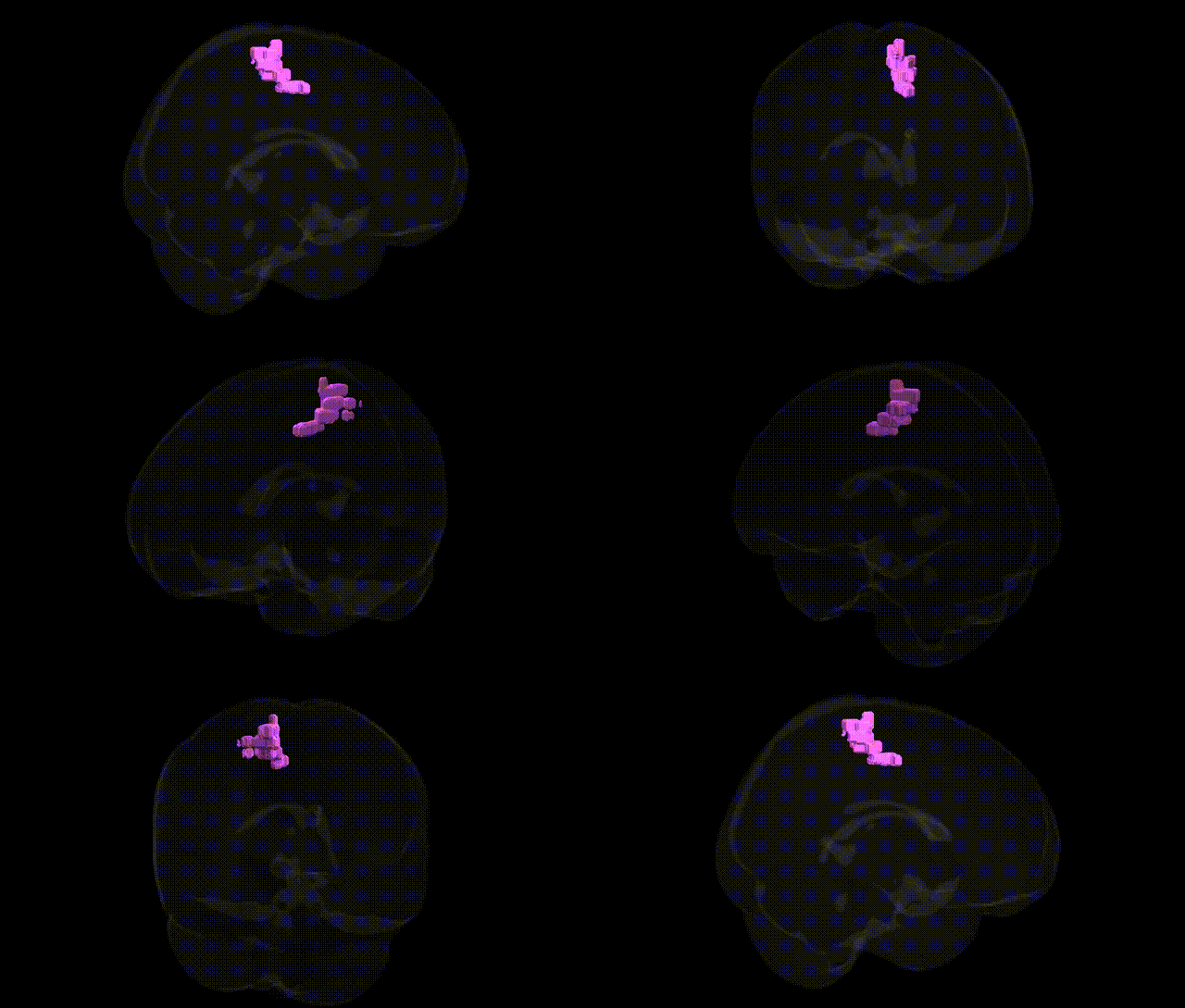
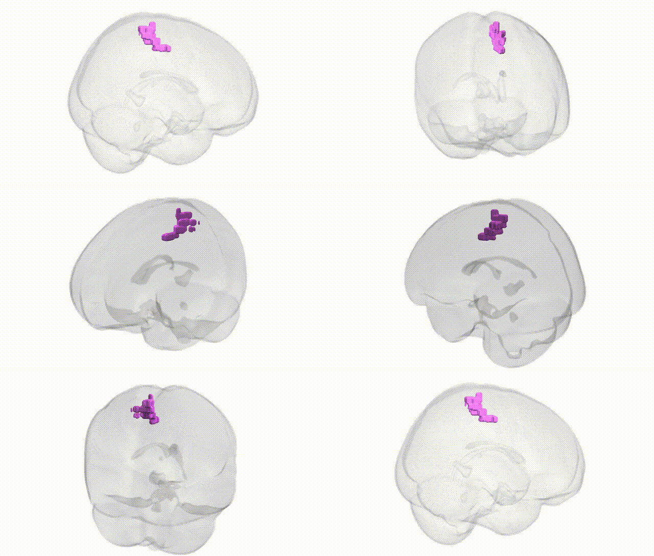
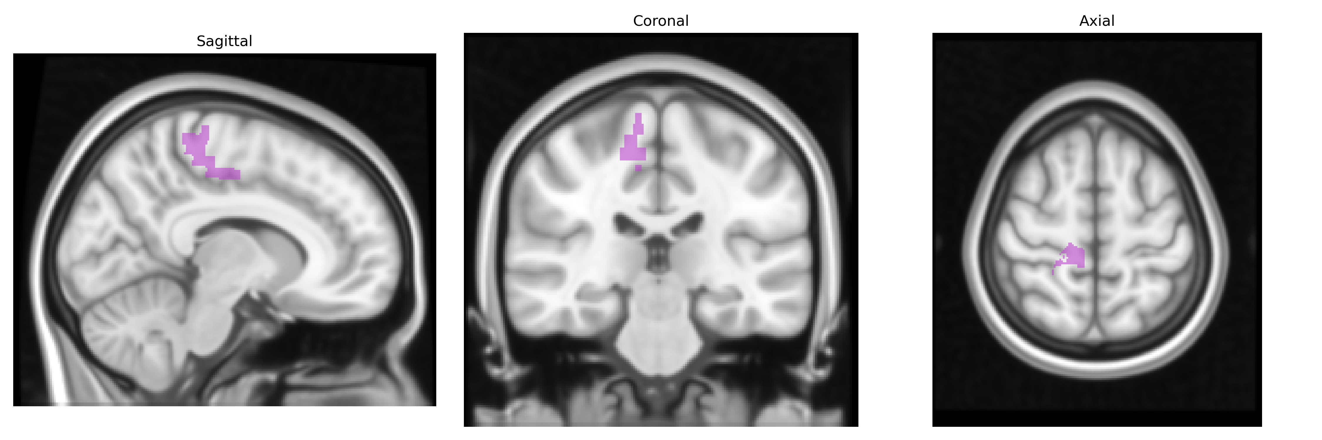
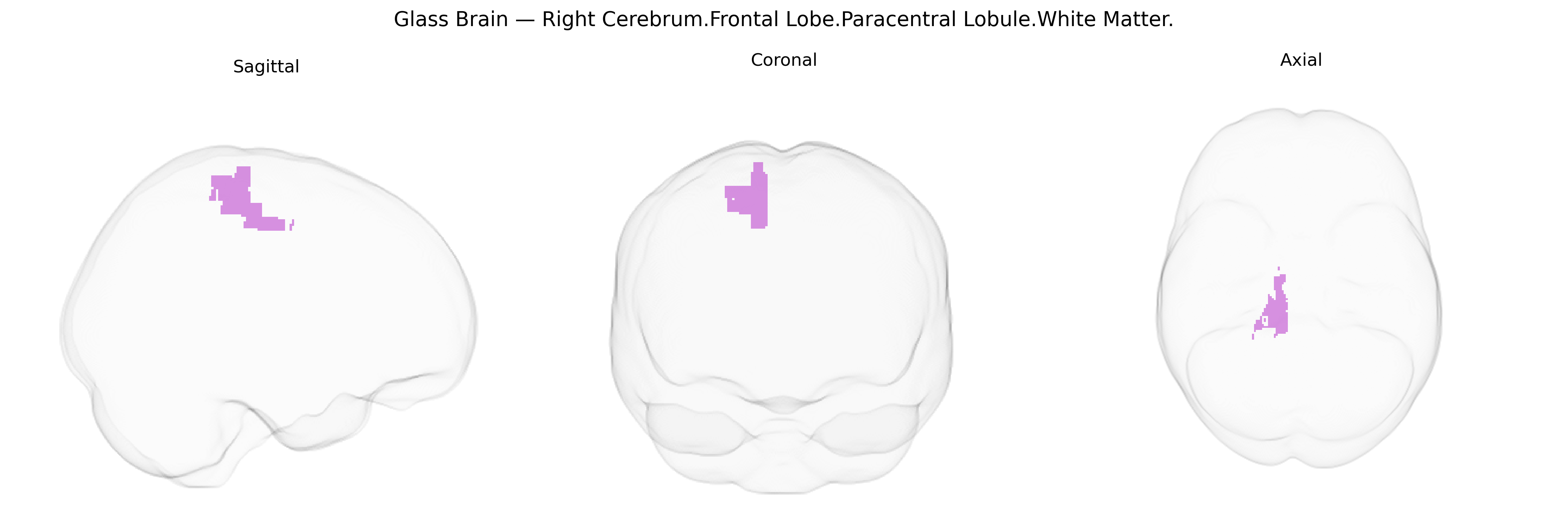
The bilateral Right Cerebrum.Frontal Lobe.Paracentral Lobule.White Matter corresponds to the deep subcortical myelinated fiber pathways underlying the paracentral lobule in the medial frontal cortex, which bridges the precentral (primary motor) and postcentral (primary somatosensory) regions around the medial continuation of the central sulcus. This white matter compartment contains short association fibers connecting adjacent medial sensorimotor cortices, as well as longer projection fibers that descend via the centrum semiovale toward the internal capsule, contributing to motor and sensory pathways controlling primarily the contralateral lower limb and perineal regions. Functionally, this region supports integration and transmission of signals related to voluntary motor control, somatosensory processing, and aspects of continence and gait. There is no direct Wikipedia article for this exact white matter label; see the related cortical region: Paracentral lobule.
The bilateral Right Cerebrum.Frontal Lobe.Paracentral Lobule.White Matter, corresponding primarily to medial primary motor and somatosensory territories for lower limbs, has been implicated in multiple genetic and GWAS-based associations through studies of brain structure, connectivity, and disease. Structural MRI and diffusion tensor imaging GWAS (e.g., ENIGMA, UK Biobank–based consortia) have identified common variants near genes involved in axon guidance, myelination, and synaptic plasticity (such as those in pathways including MAPT, NCAM1, and cell-adhesion or cytoskeletal genes) that influence white-matter microstructure and cortical thickness in medial frontal regions encompassing the paracentral lobule. This white-matter territory has been implicated in genetic risk for neurodegenerative and demyelinating conditions—most notably multiple sclerosis, amyotrophic lateral sclerosis, and hereditary spastic paraplegias—where variants in genes such as HLA-DRB1 (MS), C9orf72, SOD1, and other ALS risk loci, as well as SPG-related genes, are associated with lesions or degeneration in corticospinal and medial frontal motor pathways that traverse the paracentral white matter. Polygenic risk for Alzheimer’s disease, schizophrenia, bipolar disorder, major depression, and autism spectrum disorder has also been linked to subtle alterations in medial frontal and paracentral white-matter integrity, with implicated loci often mapping to synaptic, immune, and oligodendroglial/myelin-related genes. In population-scale imaging genetics, paracentral lobule white-matter volume and diffusion metrics show significant heritability and are modulated by genome-wide significant variants associated with global intracranial volume, cortical surface area, and fractional anisotropy, suggesting that this region participates in broad, polygenic architecture of brain morphometry and connectivity.
Overview generated by GPT-4o (2026).
Region ID: 1052
Hemisphere: bilateral
Atlas: Talairach labels 1mm
Full Quality Version: Download MP4
Full Quality Version: Download MP4
Wali Sidiqyar*, Gaurav Rudravaram*, Elyssa M. McMaster, Trent M. Schwartz, Adam M. Saunders, Kurt G. Schilling, Bennett A. Landman "Introducing SPINS: A Shared Public Visualization Library of Neuroanatomical Structures." Medical Imaging with Deep Learning- short paper
This resource is licensed under CC0 1.0 Universal (Public Domain).