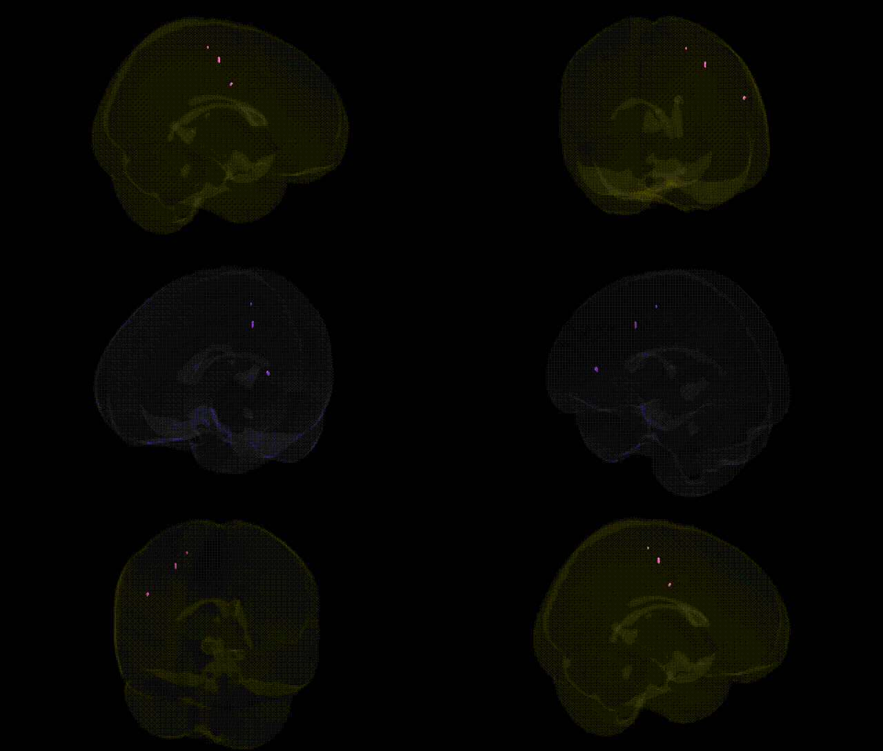
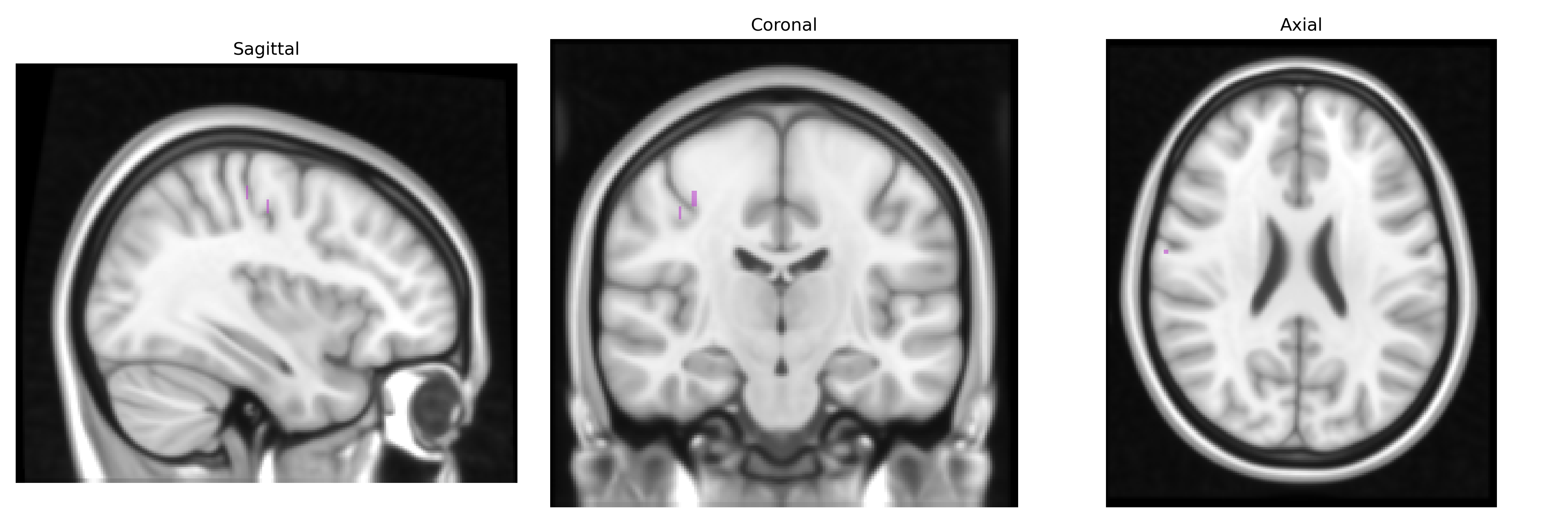
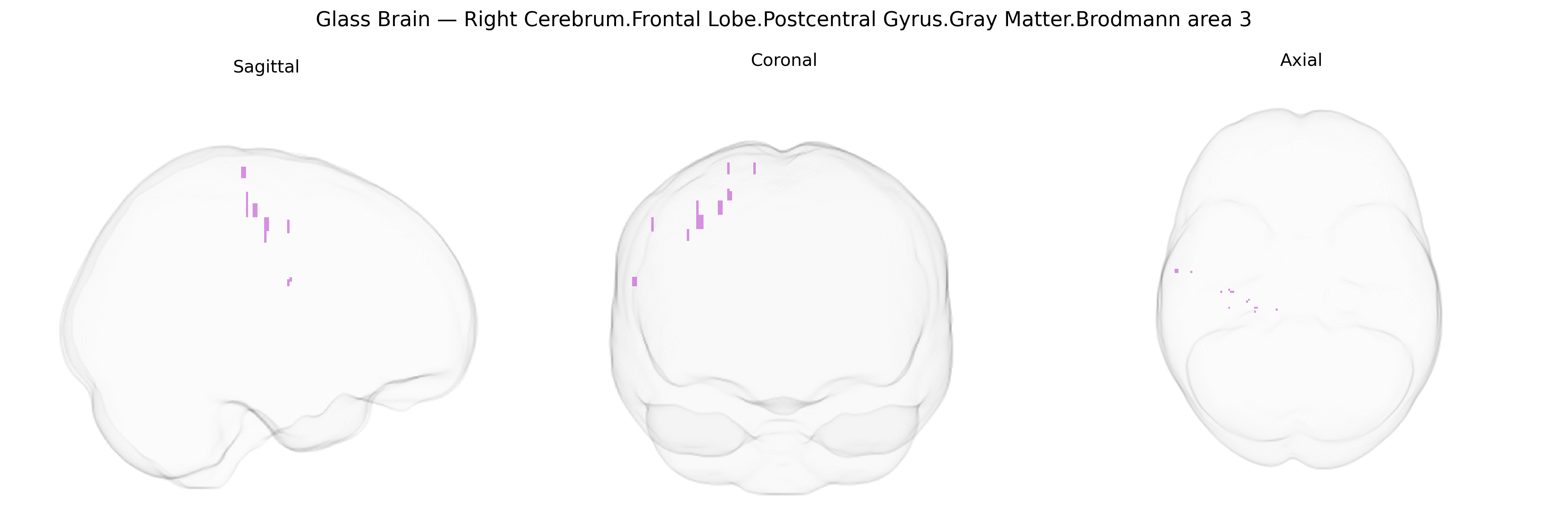
The bilateral Right Cerebrum.Frontal Lobe.Postcentral Gyrus.Gray Matter.Brodmann area 3 corresponds primarily to the primary somatosensory cortex (S1), specifically the granular cortical field located immediately posterior to the central sulcus along the postcentral gyrus. Brodmann area 3 is heavily interconnected with thalamic ventral posterior nuclei and is characterized by dense granular cell layers (notably layer IV), supporting high-resolution processing of cutaneous and proprioceptive inputs from contralateral body regions. Functionally, it is considered the earliest cortical stage of somatosensory processing, receiving finely topographic afferent projections that contribute to the somatotopic “homunculus.” Although Talairach naming here refers to the frontal lobe, this region is classically assigned to the parietal lobe. There is no direct Wikipedia article for this exact composite label; a closely related structure is the Primary somatosensory cortex.
Genetic associations involving the bilateral Right Cerebrum Frontal Lobe Postcentral Gyrus Gray Matter Brodmann area 3 (a primary somatosensory cortex subregion, as defined in the Talairach 1 mm atlas) largely mirror those reported for primary somatosensory/postcentral cortex more broadly, rather than this exact microanatomical label. GWAS and imaging-genetics studies have linked common variants in genes involved in neurodevelopment and synaptic function (for example, variants near or within genes such as BDNF, GRIN2B, and multiple axon guidance and cytoskeletal genes) to cortical thickness, surface area, and gyrification of postcentral/somatosensory regions, implicating this area in the heritable architecture of sensorimotor cortex. Large-scale consortia such as ENIGMA and UK Biobank have reported significant SNP-based heritability for postcentral gyrus morphometry, with loci in pathways related to neuronal differentiation, calcium signaling, and myelination contributing to individual differences. Disorder-focused genetic imaging studies have associated altered structure or activation of primary somatosensory cortex—including BA3—with polygenic risk for autism spectrum disorder, schizophrenia, ADHD, and developmental coordination or motor disorders, often in the context of sensory processing abnormalities or altered sensorimotor integration. Additional GWAS of chronic pain, touch sensitivity, and sensorimotor traits (e.g., grip strength, fine motor performance) have identified risk loci whose effects partially localize to somatosensory cortex, suggesting a genetic contribution to BA3-related phenotypes, although current evidence remains coarse-grained and typically does not isolate the bilateral right BA3 label specifically.
Overview generated by GPT-4o (2026).
Region ID: 898
Hemisphere: bilateral
Atlas: Talairach labels 1mm
Full Quality Version: Download MP4
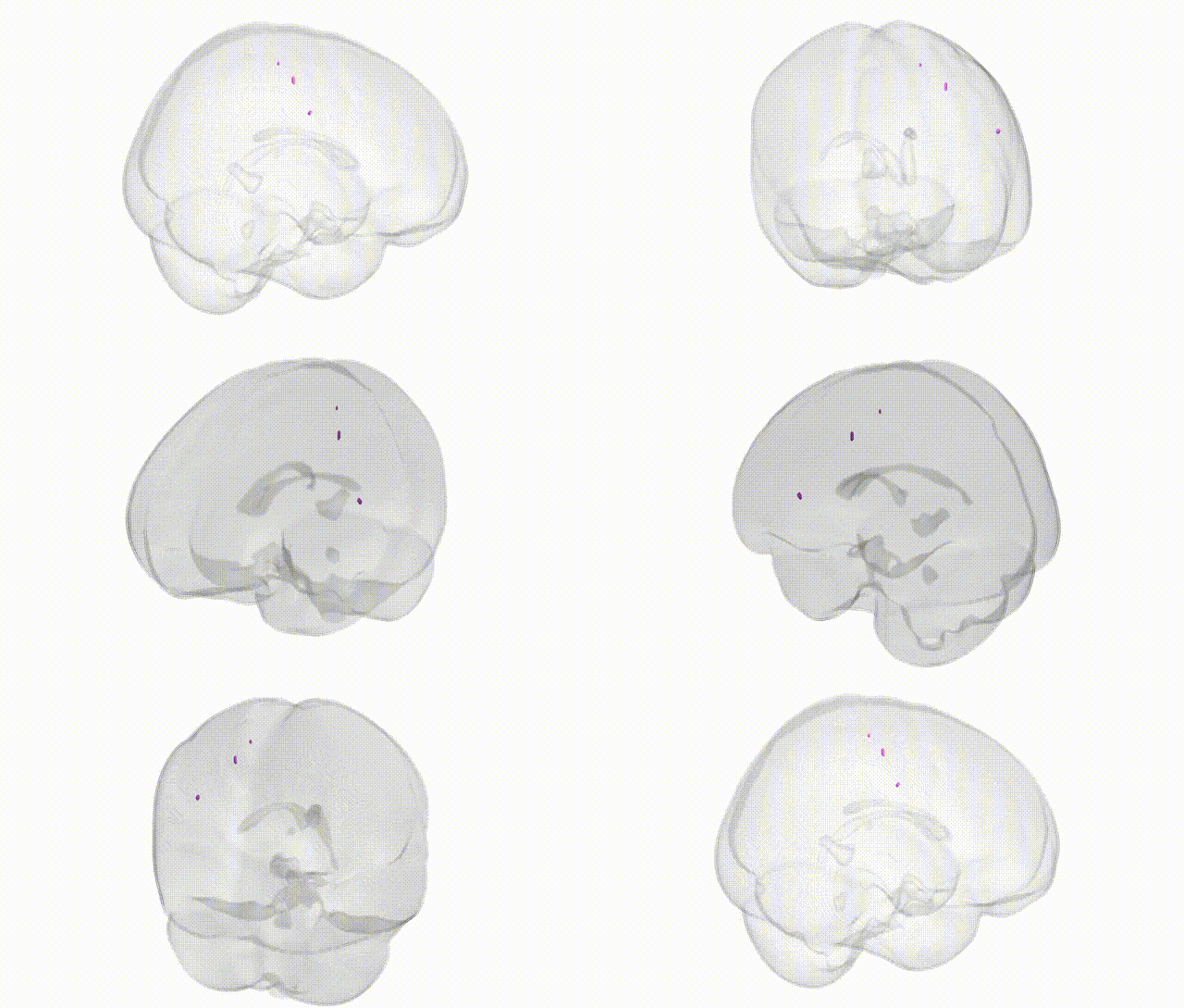
Full Quality Version: Download MP4
Wali Sidiqyar*, Gaurav Rudravaram*, Elyssa M. McMaster, Trent M. Schwartz, Adam M. Saunders, Kurt G. Schilling, Bennett A. Landman "Introducing SPINS: A Shared Public Visualization Library of Neuroanatomical Structures." Medical Imaging with Deep Learning- short paper
This resource is licensed under CC0 1.0 Universal (Public Domain).