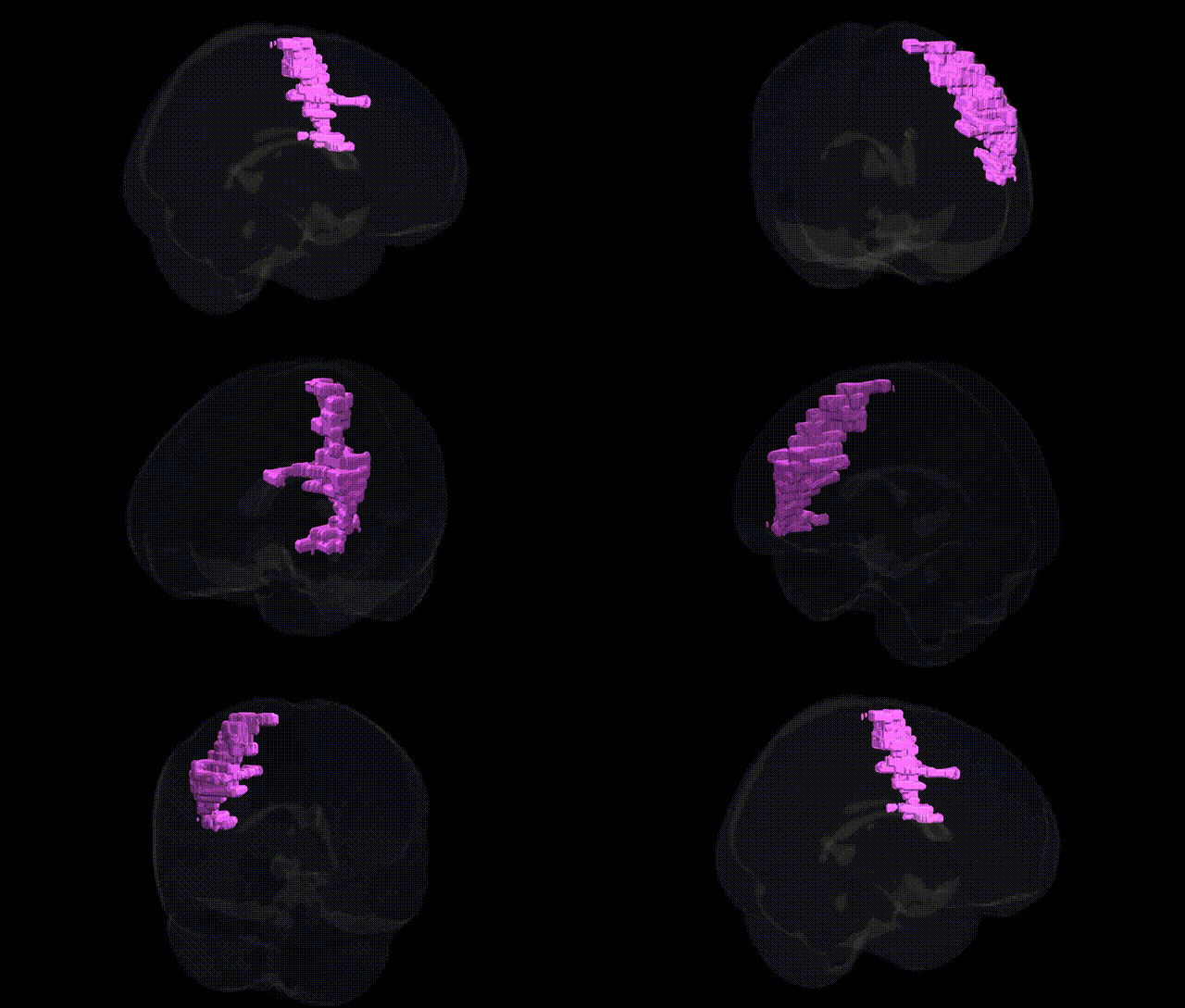
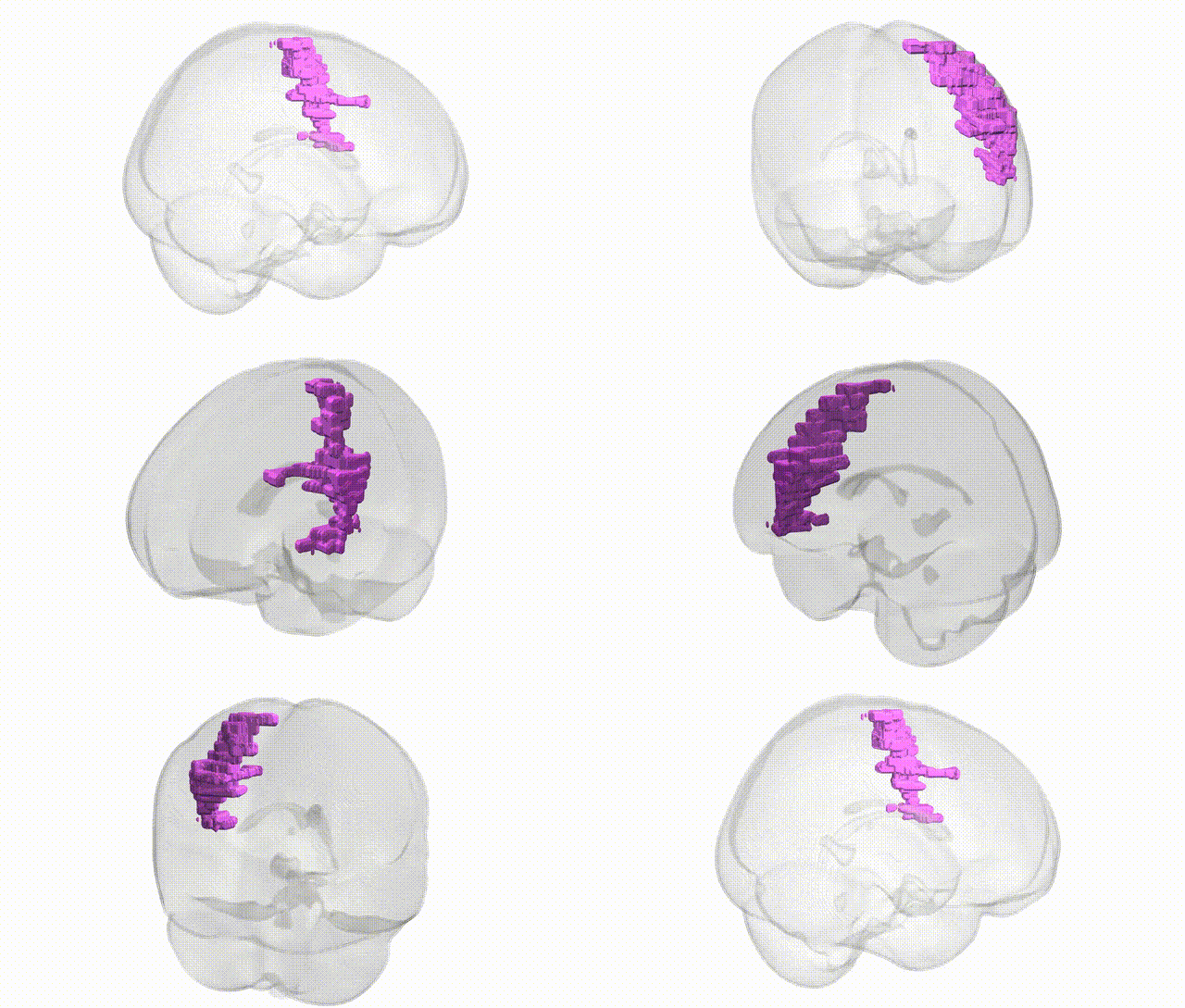
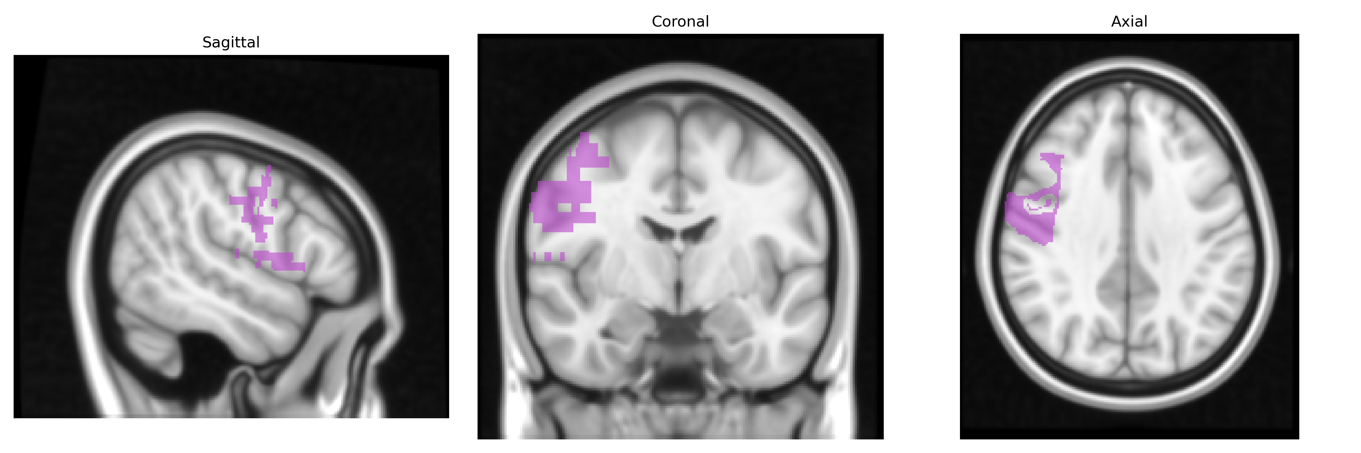
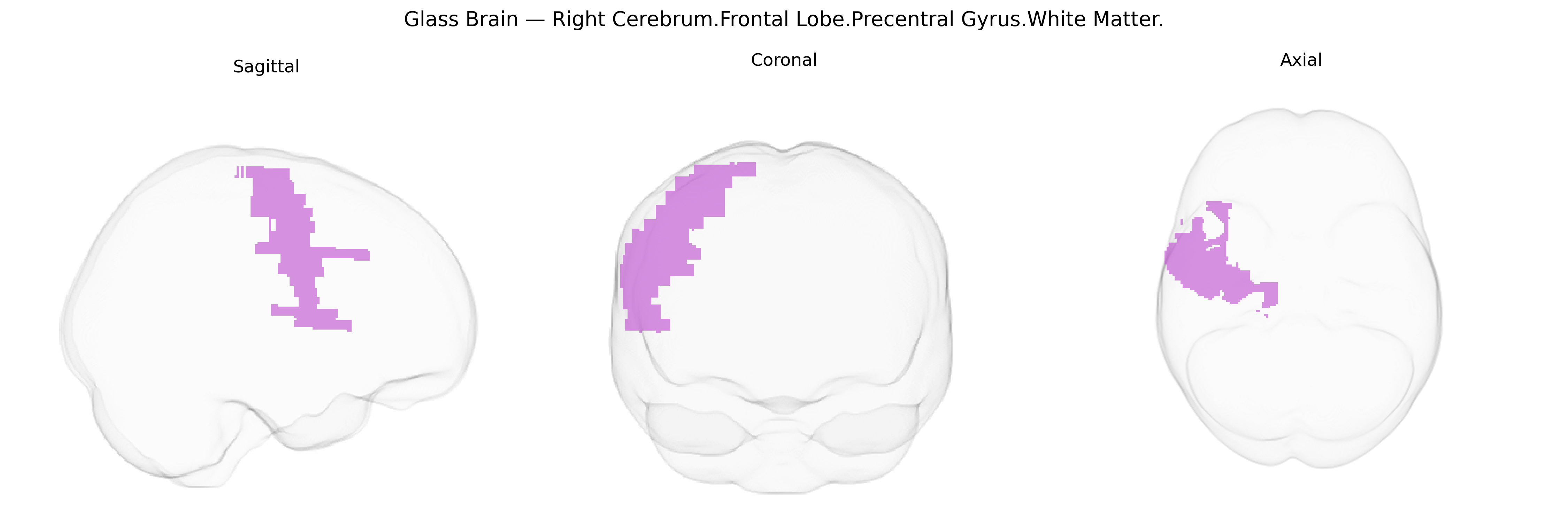
The bilateral Right Cerebrum.Frontal Lobe.Precentral Gyrus.White Matter corresponds to the deep myelinated fiber tracts underlying the right precentral gyrus, the primary motor cortex region of the frontal lobe. These white matter pathways contain corticospinal, corticobulbar, and association fibers that transmit efferent motor commands from upper motor neurons in the precentral gray matter to lower motor neurons in the brainstem and spinal cord, and also interconnect motor and premotor regions within the frontal lobe and with other cortical areas. Functionally, this white matter supports the execution and coordination of voluntary movements, somatotopically organized along the precentral gyrus, including fine motor control of the face, upper limb, and trunk, with a particular role in contralateral (left-sided) motor function due to decussation of many motor pathways in the brainstem. Precentral gyrus
The bilateral Right Cerebrum.Frontal Lobe.Precentral Gyrus.White Matter region corresponds primarily to the white-matter tracts connecting the primary motor cortex (precentral gyrus) with subcortical and spinal motor pathways, and genetic associations for this area are usually derived from imaging genetics studies of cortical thickness, surface area, and diffusion MRI measures (e.g., fractional anisotropy) rather than Talairach labels specifically. Large GWAS of brain structure (such as ENIGMA and UK Biobank-based studies) have identified numerous loci affecting frontal lobe and precentral gyrus morphology and connectivity, frequently implicating genes involved in neurodevelopment, axon guidance, and myelination (for example, variants near genes such as CNTNAP2, DISC1, MAPT, FOXP2, and myelin-related genes like MAG and MBP in broader frontal/motor circuitry). Alterations in precentral gyrus white matter have been reported in genetic studies of motor and neurodevelopmental disorders, including amyotrophic lateral sclerosis (with risk loci such as C9orf72, SOD1, TARDBP influencing corticospinal tract integrity), hereditary spastic paraplegia (SPAST, ATL1, and related genes affecting long motor tracts), and some forms of cerebral palsy with identified genetic contributors to corticospinal development. Imaging-genetic work in schizophrenia, bipolar disorder, autism spectrum disorder, and ADHD has linked risk variants (e.g., in CACNA1C, ANK3, NRXN1, and others) to altered frontal white-matter microstructure, sometimes including precentral regions as part of broader frontal motor networks. In addition, polygenic scores for general cognitive ability, educational attainment, and risk-taking behavior have been associated with variation in frontal white matter integrity and volume in large cohorts, suggesting that many common variants with small effects jointly shape the structural and functional properties of this motor-frontal white matter zone.
Overview generated by GPT-4o (2026).
Region ID: 687
Hemisphere: bilateral
Atlas: Talairach labels 1mm
Full Quality Version: Download MP4
Full Quality Version: Download MP4
Wali Sidiqyar*, Gaurav Rudravaram*, Elyssa M. McMaster, Trent M. Schwartz, Adam M. Saunders, Kurt G. Schilling, Bennett A. Landman "Introducing SPINS: A Shared Public Visualization Library of Neuroanatomical Structures." Medical Imaging with Deep Learning- short paper
This resource is licensed under CC0 1.0 Universal (Public Domain).