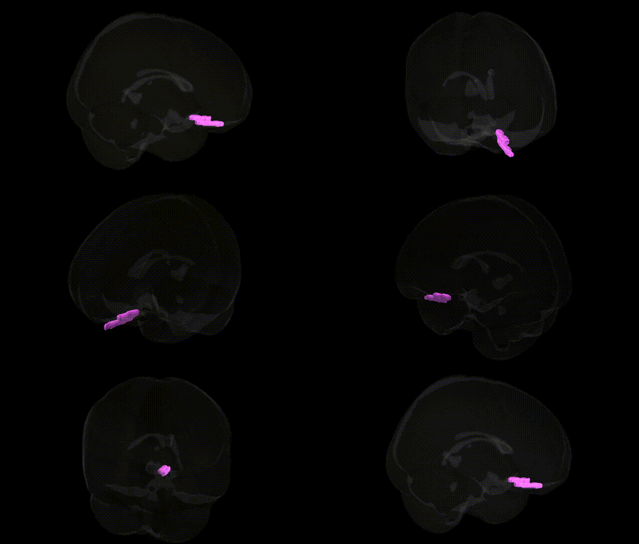
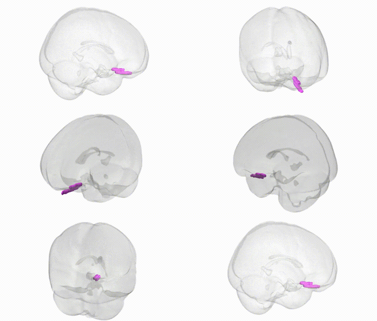
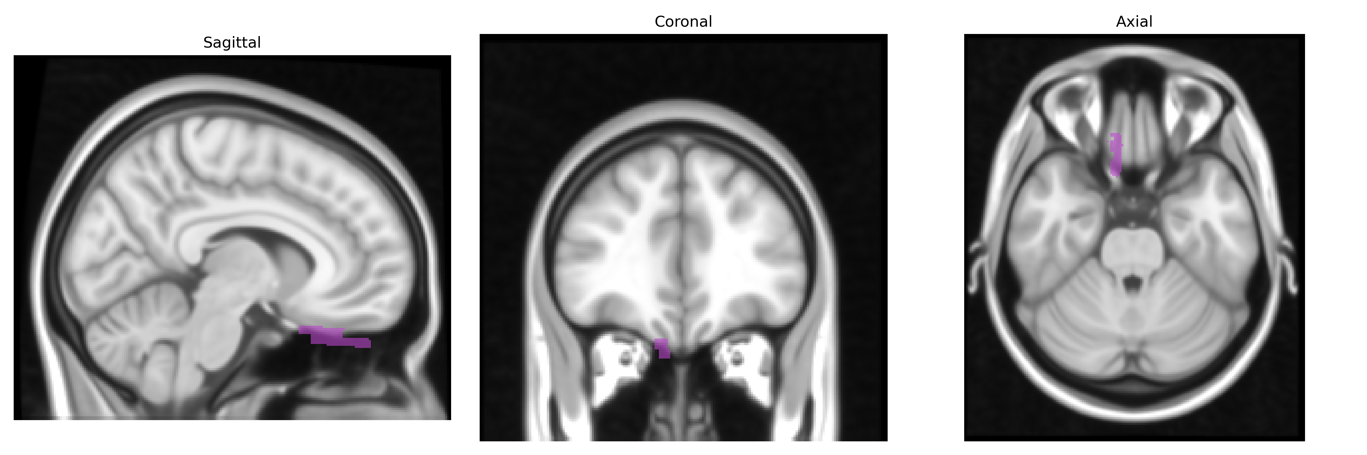
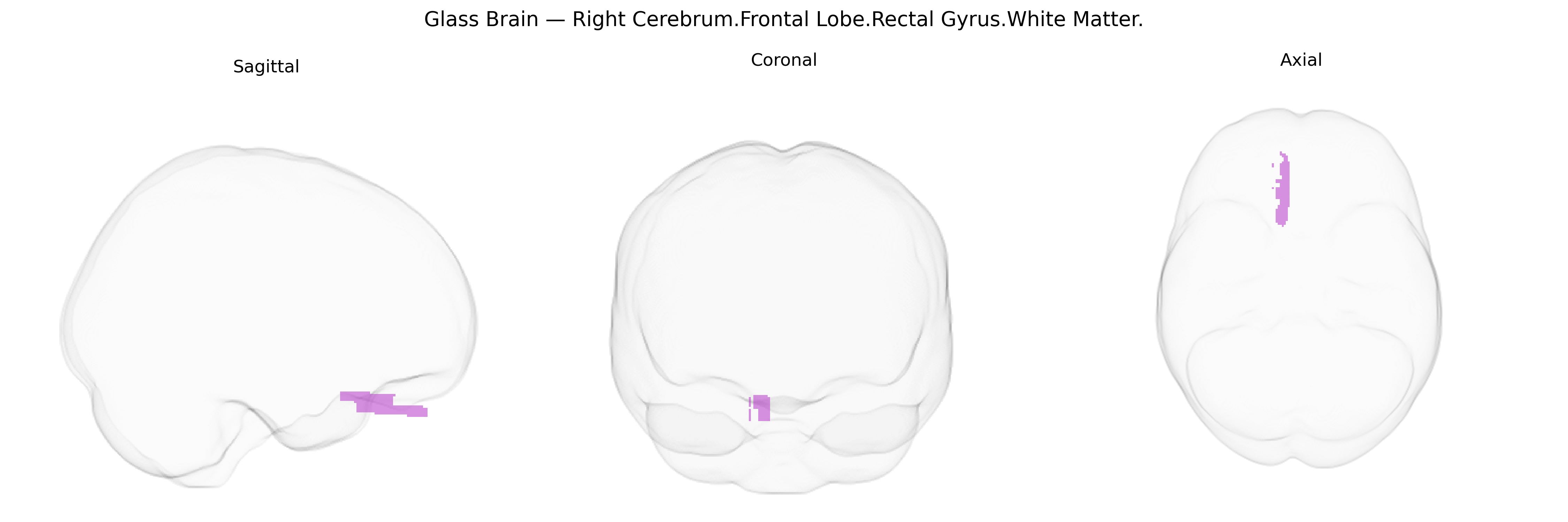
The bilateral right cerebrum frontal lobe rectal gyrus white matter corresponds to subcortical fiber pathways underlying the medial orbitofrontal cortex, particularly deep to the gyrus rectus (also called the straight gyrus) along the inferior surface of the frontal lobe adjacent to the olfactory sulcus and frontal pole. These white matter tracts participate in fronto-limbic and fronto-basal connections that support reward evaluation, affective processing, decision-making, and aspects of social behavior, integrating input from limbic structures (such as the amygdala and hypothalamus) and projecting to other prefrontal and subcortical regions. Because this is a white matter designation in the Talairach 1 mm atlas rather than a distinct named fasciculus, it is functionally interpreted in the context of the overlying medial orbitofrontal and gyrus rectus gray matter. There is no direct link for this precise white matter region; a related cortical area is the Orbitofrontal cortex.
The bilateral right rectal (gyrus rectus) white matter of the frontal lobe, as defined in the Talairach 1 mm atlas, has not been a primary target in most genetic studies, but convergent evidence from imaging-genetics and GWAS implicates nearby orbitofrontal/medial frontal white matter and adjacent gyrus rectus in several traits and disorders. Large neuroimaging GWAS consortia (e.g., ENIGMA, UK Biobank) have identified common variants in genes related to axonal growth, myelination, and synaptic function (such as BDNF, NTRK2, CNTNAP2, and various cell-adhesion and guidance genes) that modulate frontal and orbitofrontal white-matter microstructure, often measured by diffusion MRI metrics like fractional anisotropy. These genetic influences on orbitofrontal–medial frontal white matter have been linked to risk for major depressive disorder, bipolar disorder, schizophrenia, and obsessive–compulsive and anxiety-related traits, as well as neuroticism, risk-taking, and cognitive control measures, consistent with the role of the orbitofrontal/ventromedial prefrontal region in affective and reward processing. Some Alzheimer’s disease and general cognitive-function GWAS also show associations with frontal white-matter integrity that include or border the gyrus rectus region, although these findings typically report larger lobar or tract-based regions (e.g., anterior cingulum, uncinate fasciculus) rather than the rectal gyrus white matter specifically, so genetic associations with this exact atlas label are inferred from broader orbitofrontal and medial frontal white-matter findings rather than directly established.
Overview generated by GPT-4o (2026).
Region ID: 149
Hemisphere: bilateral
Atlas: Talairach labels 1mm
Full Quality Version: Download MP4
Full Quality Version: Download MP4
Wali Sidiqyar*, Gaurav Rudravaram*, Elyssa M. McMaster, Trent M. Schwartz, Adam M. Saunders, Kurt G. Schilling, Bennett A. Landman "Introducing SPINS: A Shared Public Visualization Library of Neuroanatomical Structures." Medical Imaging with Deep Learning- short paper
This resource is licensed under CC0 1.0 Universal (Public Domain).