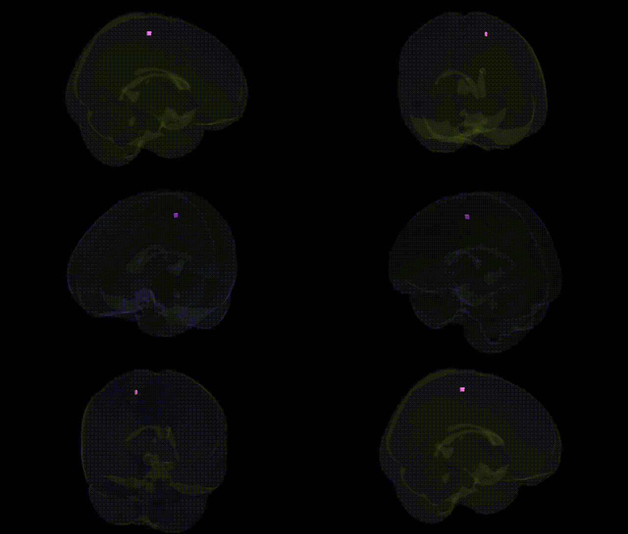
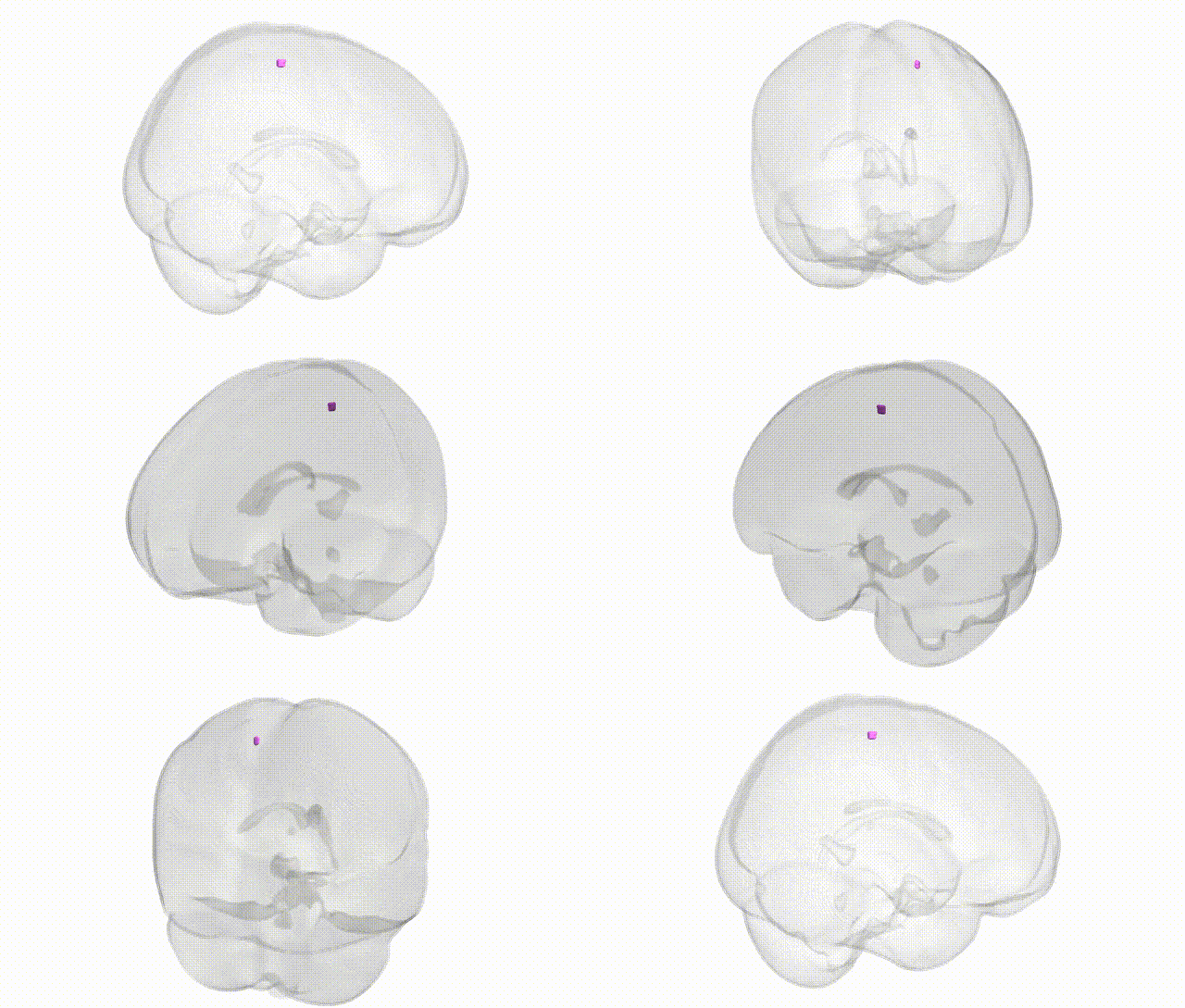
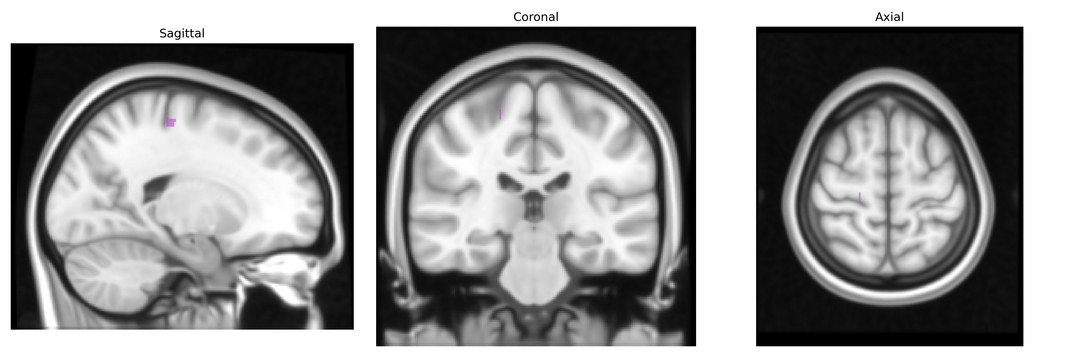
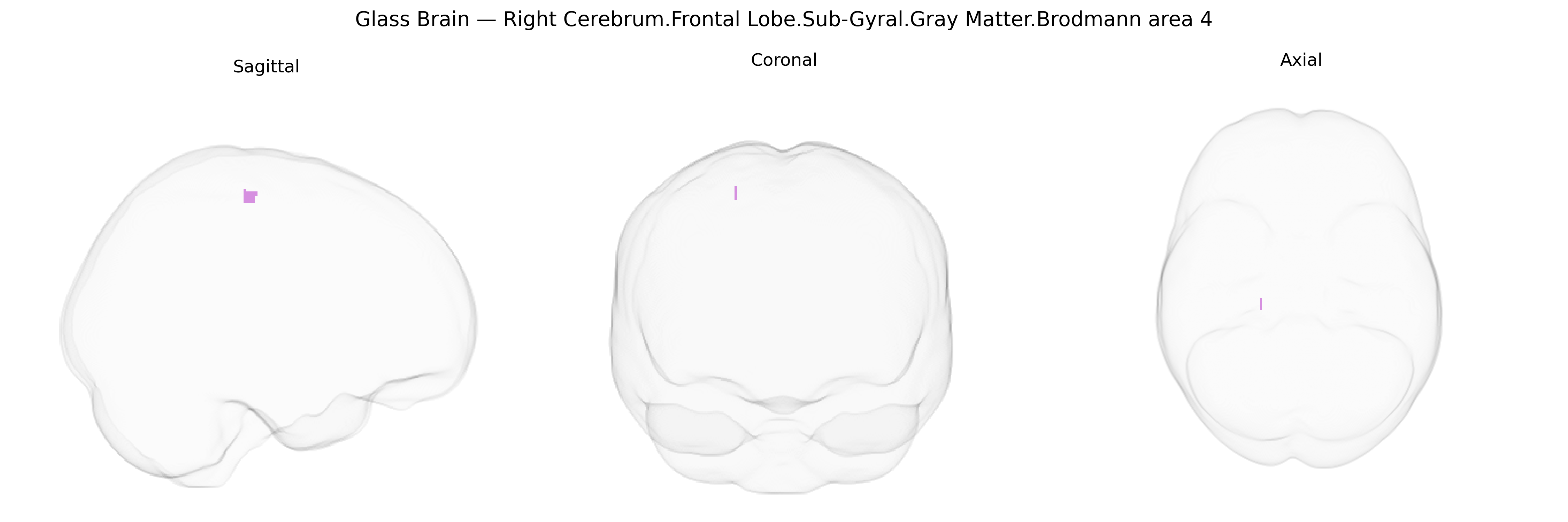
The bilateral Right Cerebrum.Frontal Lobe.Sub-Gyral.Gray Matter.Brodmann area 4 corresponds primarily to the primary motor cortex located in the precentral gyrus, deep to the cortical surface within subgyral gray matter of the frontal lobe. This agranular cortical area contains large pyramidal (Betz) cells projecting via the corticospinal and corticobulbar tracts to spinal and brainstem motor nuclei, thereby mediating voluntary execution of fine, goal-directed movements, particularly of the contralateral body. Somatotopic organization within this region forms part of the motor homunculus, with distinct representations for different muscle groups and dense connectivity to premotor, supplementary motor, and somatosensory areas. A directly corresponding Wikipedia entry is not available; a closely related structure is the Primary motor cortex.
The bilateral right cerebrum frontal lobe sub-gyral gray matter corresponding to Brodmann area 4 (primary motor cortex) has been implicated in several genetic association and imaging-genetics findings, though typically reported at the level of motor cortex, precentral gyrus, or frontal lobe rather than the exact Talairach parcel. GWAS of brain structure (e.g., ENIGMA and UK Biobank) have identified common variants near genes such as HMGA2, IGF1, and others associated with global and regional cortical thickness and surface area, including motor and frontal regions encompassing BA4. Motor cortical structure and function show heritability, and variants in genes involved in neurodevelopment and synaptic function (e.g., CNTNAP2, BDNF, GRIN2B) have been linked to traits that involve BA4 circuitry, such as motor skill learning, handedness, and cortical excitability. Disorders with prominent motor involvement—Parkinson’s disease, amyotrophic lateral sclerosis, and certain hereditary spastic paraplegias—show structural and functional changes in BA4 and have robust genetic architectures (e.g., SNCA, LRRK2, GBA; SOD1, C9orf72, TARDBP; SPAST), although genetic risk is typically not specific to BA4 but affects broader motor networks. Imaging-genetics studies in autism spectrum disorder, ADHD, and schizophrenia have reported frontal and motor cortical alterations associated with risk variants (e.g., CACNA1C, MIR137, DRD2), with BA4 often part of the affected sensorimotor circuitry. Overall, genetic contributions to variation in BA4 structure and function are supported by polygenic influences on cortical morphology and motor traits, but there is limited evidence for variants that selectively and specifically target this Talairach-defined sub-gyral BA4 region.
Overview generated by GPT-4o (2026).
Region ID: 1093
Hemisphere: bilateral
Atlas: Talairach labels 1mm
Full Quality Version: Download MP4
Full Quality Version: Download MP4
Wali Sidiqyar*, Gaurav Rudravaram*, Elyssa M. McMaster, Trent M. Schwartz, Adam M. Saunders, Kurt G. Schilling, Bennett A. Landman "Introducing SPINS: A Shared Public Visualization Library of Neuroanatomical Structures." Medical Imaging with Deep Learning- short paper
This resource is licensed under CC0 1.0 Universal (Public Domain).