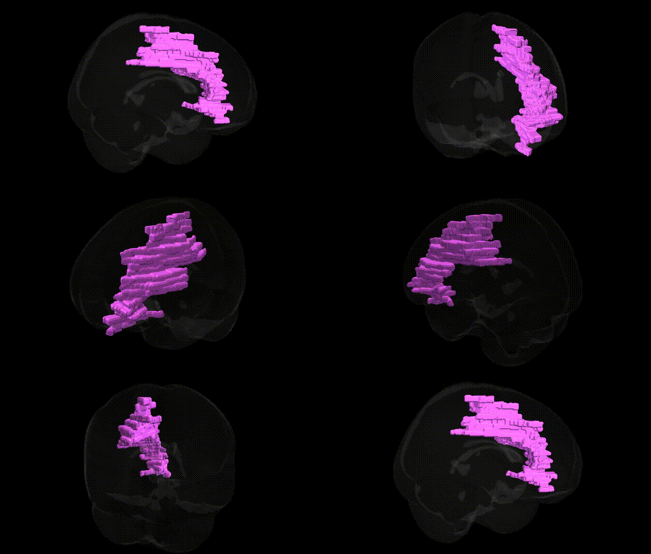
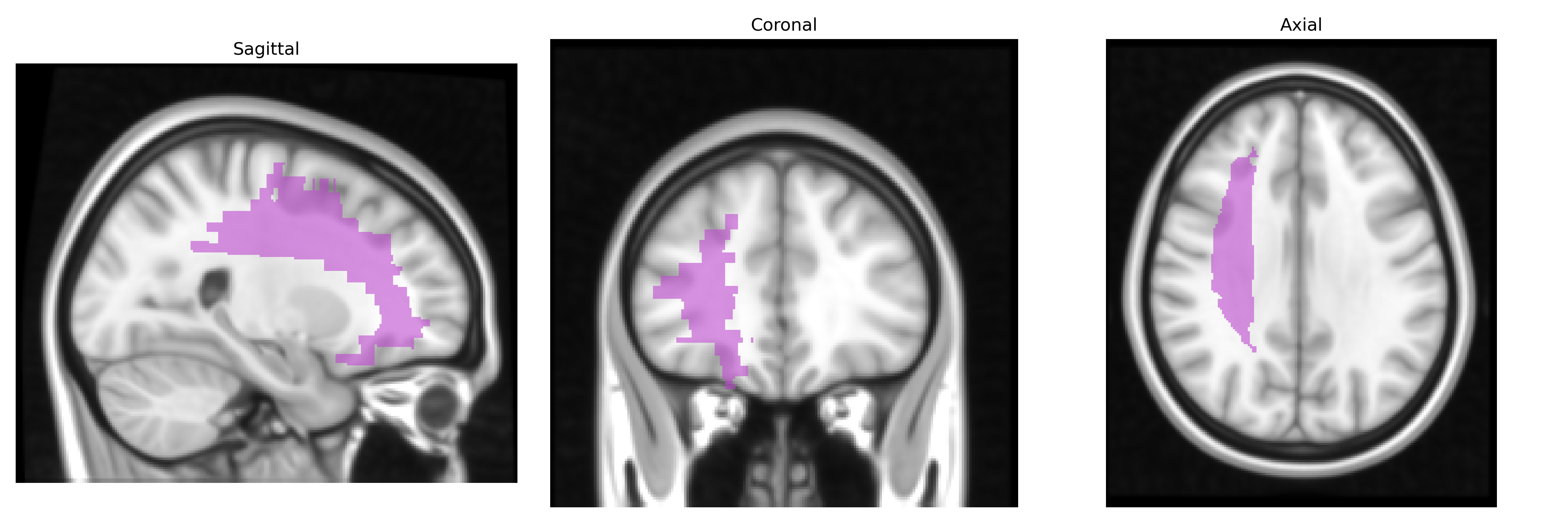
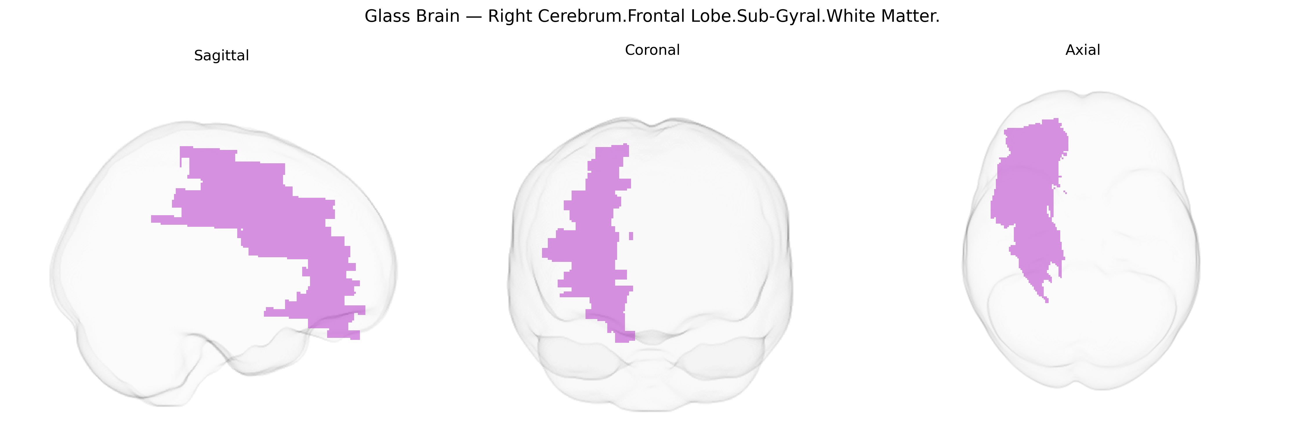
The bilateral Right Cerebrum.Frontal Lobe.Sub-Gyral.White Matter corresponds to deep frontal lobe white matter lying beneath the cortical gyri of the right frontal lobe, encompassing association and projection fibers that interconnect prefrontal, premotor, and motor cortices with each other and with subcortical structures. This region contains major frontal white matter pathways, including portions of the superior longitudinal fasciculus, fronto-striatal and fronto-thalamic projections, and short U-fibers linking adjacent gyri, supporting higher-order executive functions, working memory, attention, motor planning, and aspects of behavior and mood regulation. As an integral part of the frontal lobe white matter, it plays a key role in distributed frontal networks rather than serving a single isolated function. There is no direct link for this exact sub-gyral white matter label; see the related structure Frontal lobe.
Genetic associations with the bilateral Right Cerebrum.Frontal Lobe.Sub-Gyral.White Matter region, as defined in the Talairach 1 mm atlas, arise primarily from GWAS and imaging-genetics studies of frontal white matter volume, integrity, and connectivity rather than this exact label per se; loci in genes involved in myelination, axon guidance, and neurodevelopment—such as MAG, MBP, NRG1, NCAM1, and contactin-related genes—have been linked to variability in frontal white matter microstructure (e.g., fractional anisotropy and mean diffusivity). Large consortia (ENIGMA, UK Biobank) report that common variants near genes like VCAN, PLEKHG1, SHROOM3, and others associated with global and lobe-specific white matter volumes also implicate frontal subgyral regions, with polygenic influences overlapping those for cognitive performance, educational attainment, and general intelligence. In neuropsychiatric genetics, risk variants for schizophrenia (e.g., in CACNA1C, ZNF804A, and other GWAS loci), bipolar disorder, major depression, and ADHD have been associated with altered frontal white matter structure, including subgyral and prefrontal tracts, while genes regulating inflammatory and vascular pathways (e.g., APOE, NOTCH3, and other small-vessel disease loci) contribute to white matter hyperintensities that often involve frontal subgyral areas. Neurodegenerative disease genes such as APOE (Alzheimer’s disease) and C9orf72 or MAPT (frontotemporal dementia) are associated with frontal white matter degeneration patterns, and imaging-genetic correlation studies link these variants with reduced integrity of frontal subgyral tracts. Together, current evidence supports a highly polygenic architecture in which genes affecting oligodendrocyte function, synaptic development, vascular health, and neuroinflammation contribute to interindividual differences and disease-related alterations in this frontal subgyral white matter region.
Overview generated by GPT-4o (2026).
Region ID: 194
Hemisphere: bilateral
Atlas: Talairach labels 1mm
Full Quality Version: Download MP4
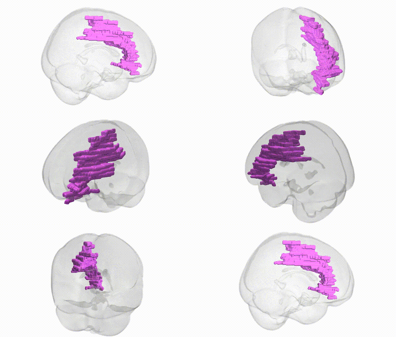
Full Quality Version: Download MP4
Wali Sidiqyar*, Gaurav Rudravaram*, Elyssa M. McMaster, Trent M. Schwartz, Adam M. Saunders, Kurt G. Schilling, Bennett A. Landman "Introducing SPINS: A Shared Public Visualization Library of Neuroanatomical Structures." Medical Imaging with Deep Learning- short paper
This resource is licensed under CC0 1.0 Universal (Public Domain).