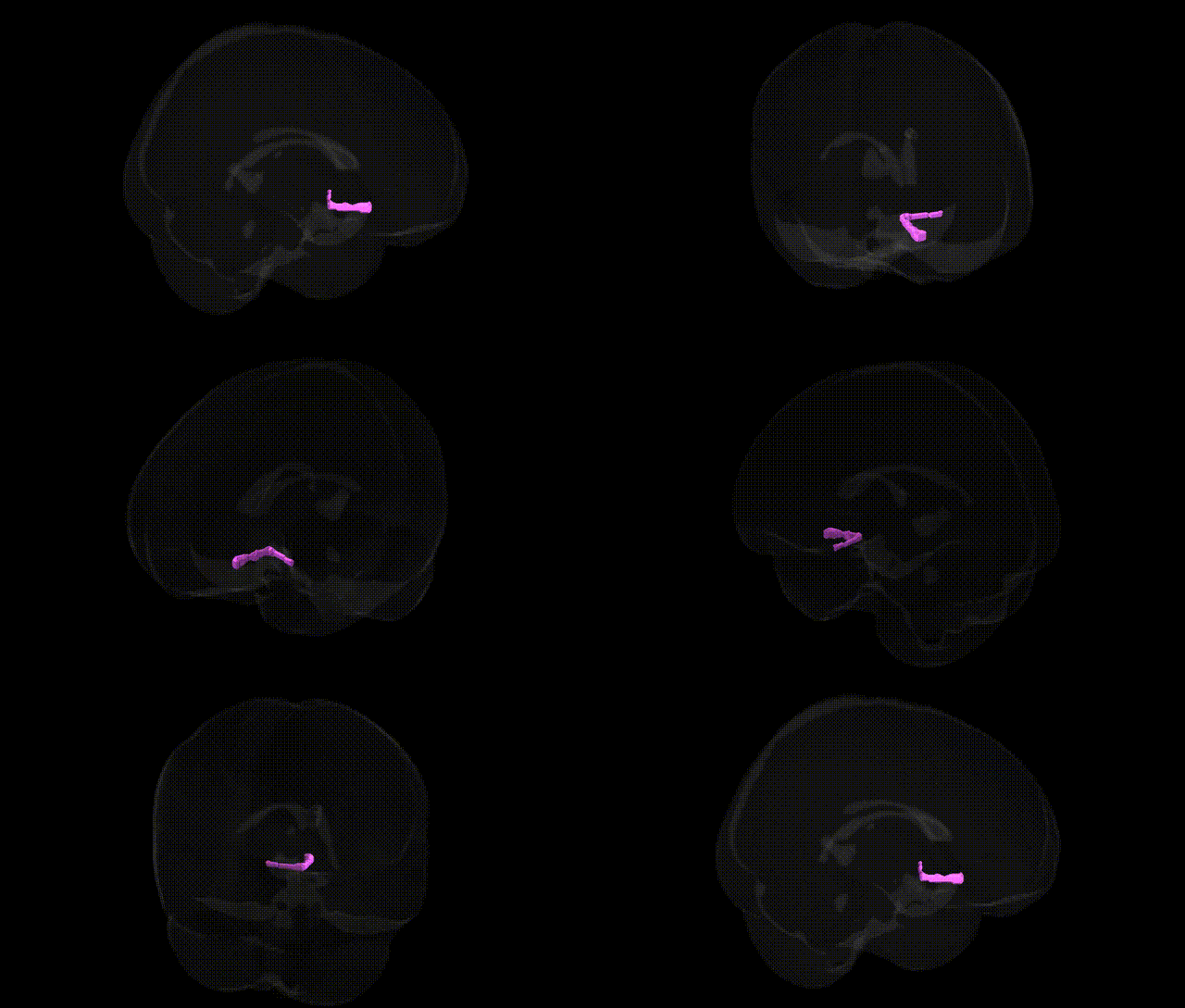
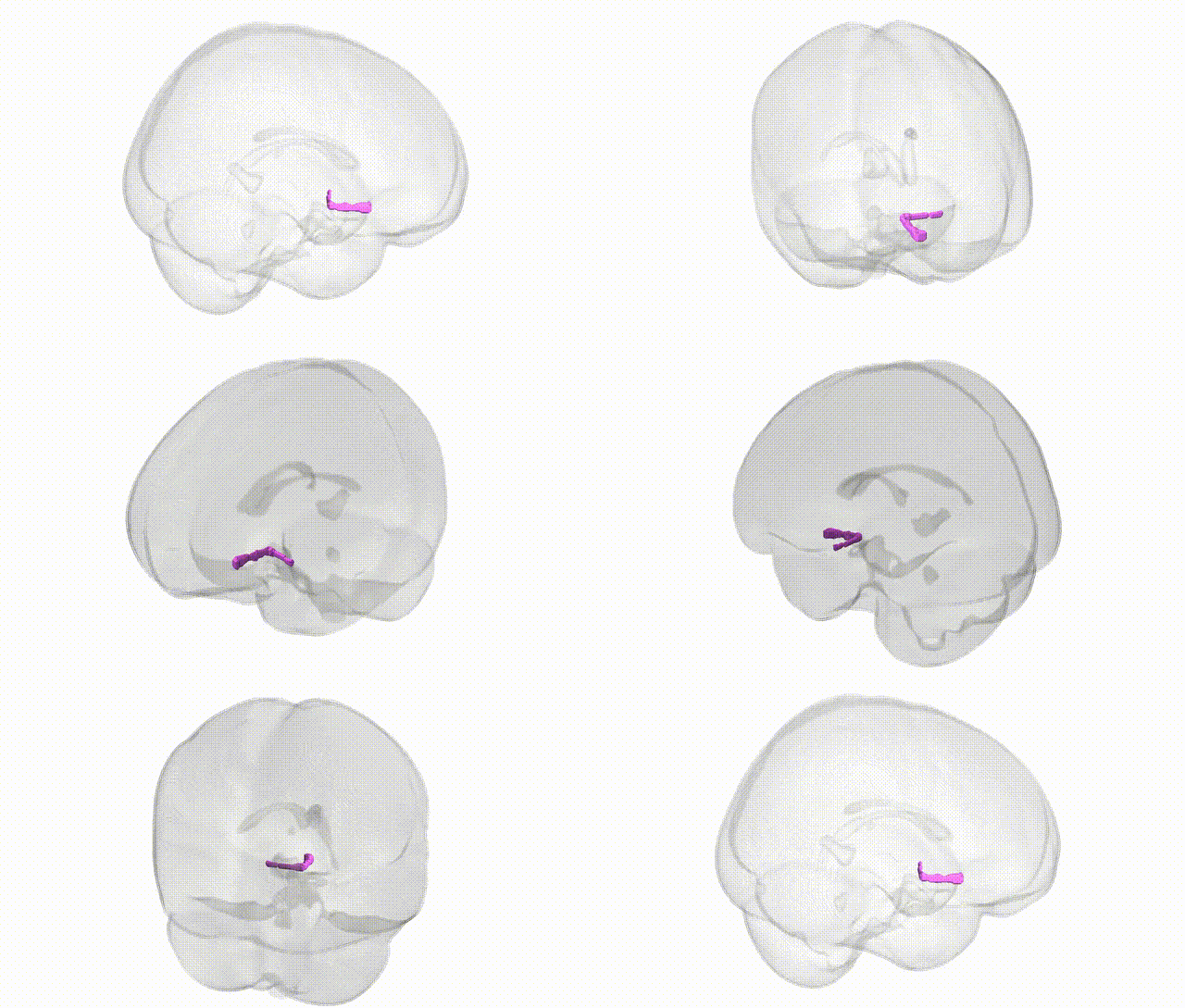
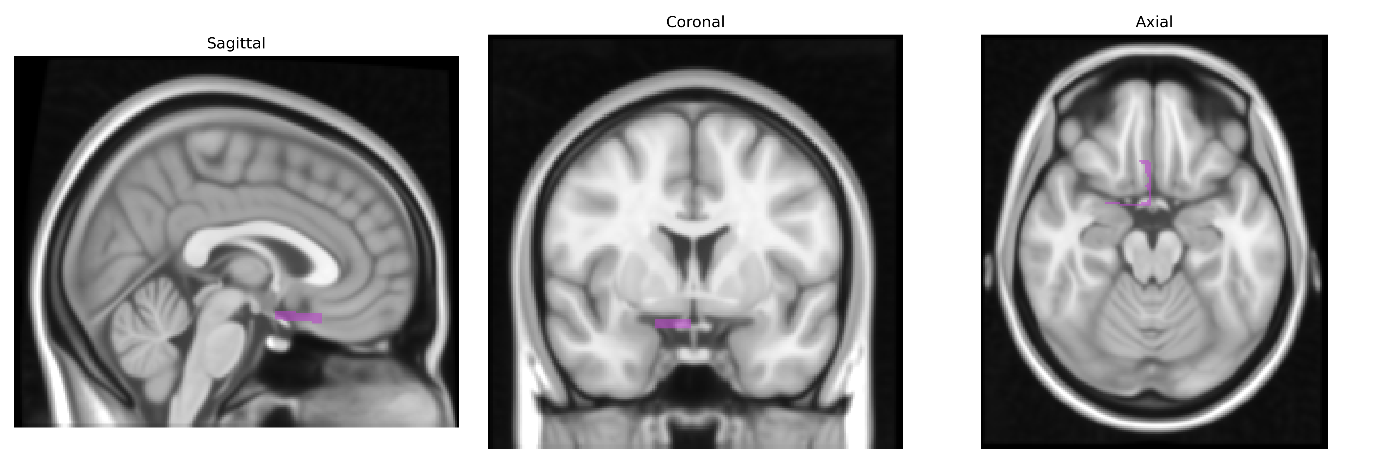
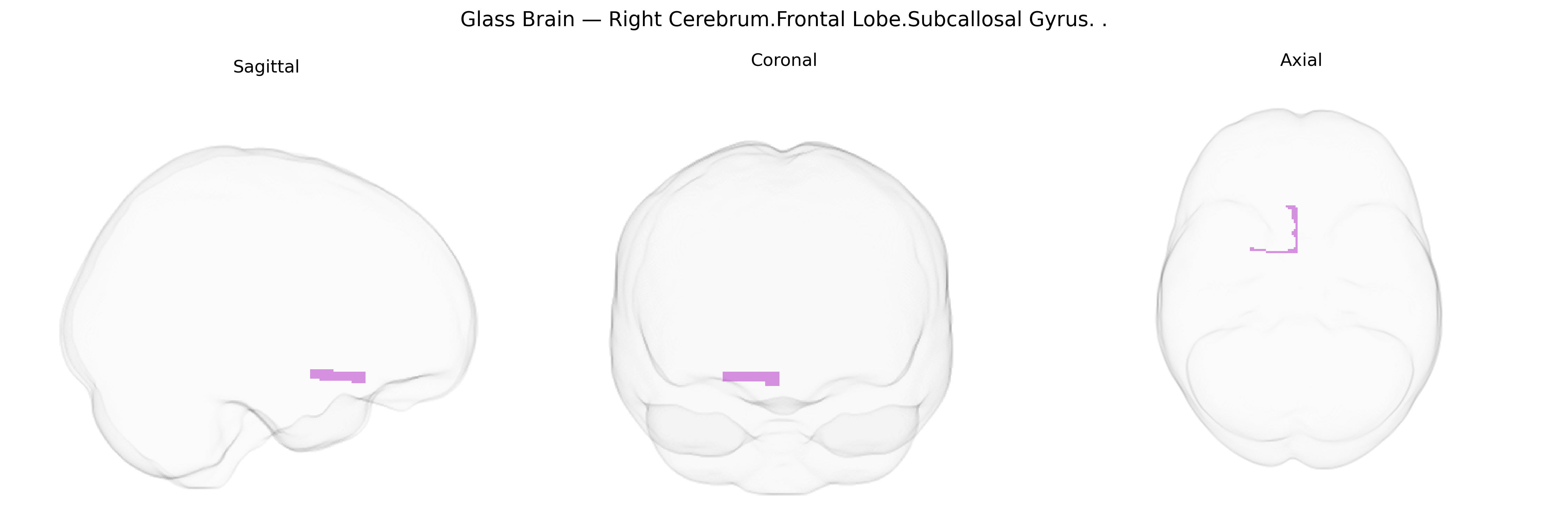
The bilateral Right Cerebrum.Frontal Lobe.Subcallosal Gyrus corresponds to the subcallosal area (also known as Brodmann area 25 or the subgenual cingulate cortex), a ventral medial prefrontal region located beneath the genu of the corpus callosum at the base of the frontal lobe. This region is part of the limbic–cortical network and is heavily interconnected with the hypothalamus, amygdala, hippocampus, and other medial prefrontal and cingulate areas, supporting roles in emotional processing, mood regulation, autonomic control, and visceromotor responses. It is notable for its involvement in major depressive disorder and treatment-resistant depression, where abnormal activity and connectivity have been reported, and it has been a target for deep brain stimulation interventions. There is no direct link; a related structure is the Subgenual anterior cingulate.
The bilateral right subcallosal gyrus (Right Cerebrum.Frontal Lobe.Subcallosal Gyrus in the Talairach 1 mm atlas), typically mapped to ventromedial prefrontal / subgenual anterior cingulate cortex (often Brodmann area 25), has been repeatedly implicated in genetic studies of mood and affective disorders, particularly major depressive disorder (MDD) and bipolar disorder, as well as traits related to negative affect and neuroticism. Structural and functional imaging–genetics work shows that common variants in serotonin transporter genes (e.g., SLC6A4), BDNF, and several stress- and synapse-related genes modulate subcallosal volume, activity, and connectivity, often in the context of depression risk, antidepressant response, and susceptibility to stress. Large-scale GWAS and imaging-genetics consortia (e.g., ENIGMA, UK Biobank analyses) have linked polygenic risk scores for MDD, bipolar disorder, and schizophrenia to altered cortical thickness or surface area in ventromedial prefrontal / subcallosal regions, and some loci associated with these disorders (for example in CACNA1C, ODZ4, and other neurodevelopmental or synaptic genes) show expression patterns enriched in frontal limbic territories including the subcallosal gyrus. Additional GWAS of personality traits, emotion regulation, and autonomic stress responses suggest that genetic variation influencing fronto-limbic circuitry, encompassing the subcallosal gyrus, contributes to individual differences in mood, anxiety-related traits, and treatment outcomes in depression, although few variants map uniquely to this region and most effects appear polygenic and distributed across broader frontal–limbic networks.
Overview generated by GPT-4o (2026).
Region ID: 278
Hemisphere: bilateral
Atlas: Talairach labels 1mm
Full Quality Version: Download MP4
Full Quality Version: Download MP4
Wali Sidiqyar*, Gaurav Rudravaram*, Elyssa M. McMaster, Trent M. Schwartz, Adam M. Saunders, Kurt G. Schilling, Bennett A. Landman "Introducing SPINS: A Shared Public Visualization Library of Neuroanatomical Structures." Medical Imaging with Deep Learning- short paper
This resource is licensed under CC0 1.0 Universal (Public Domain).