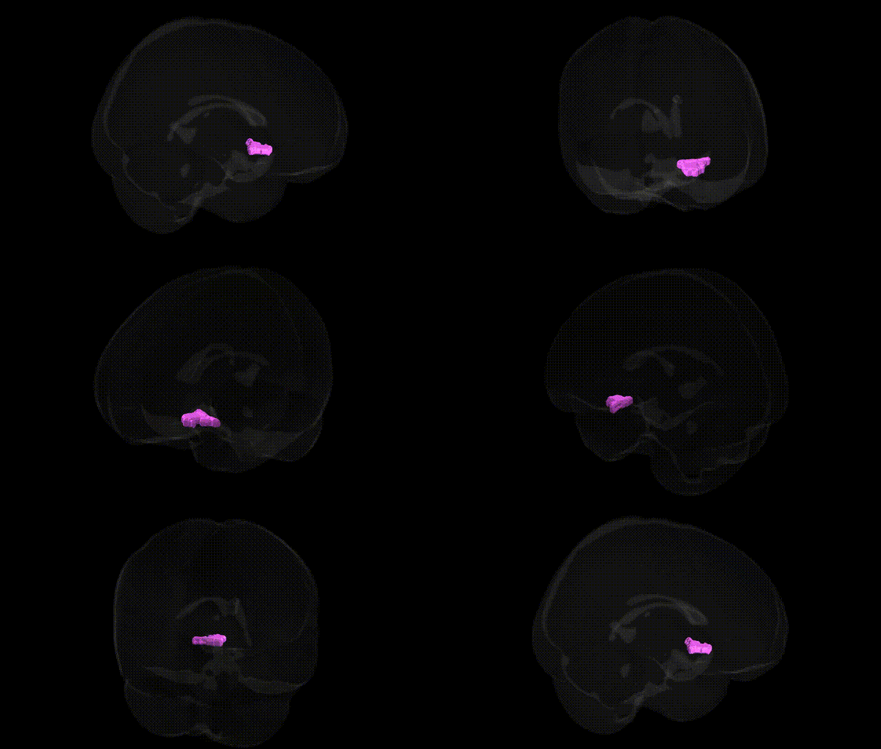
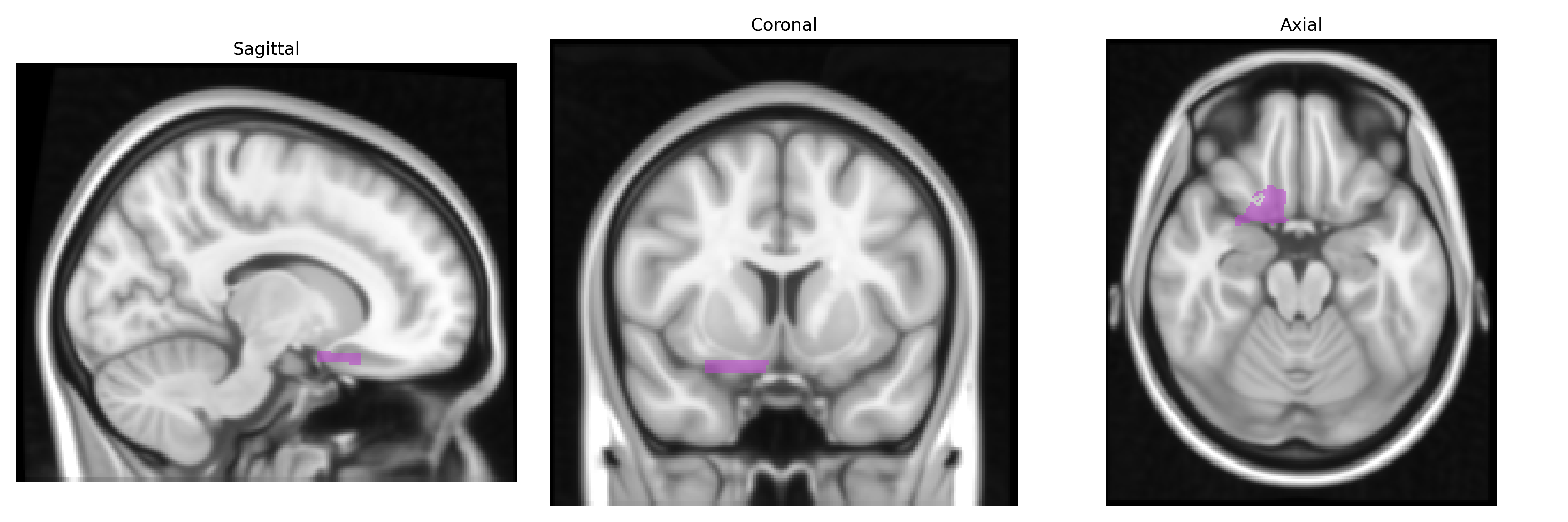
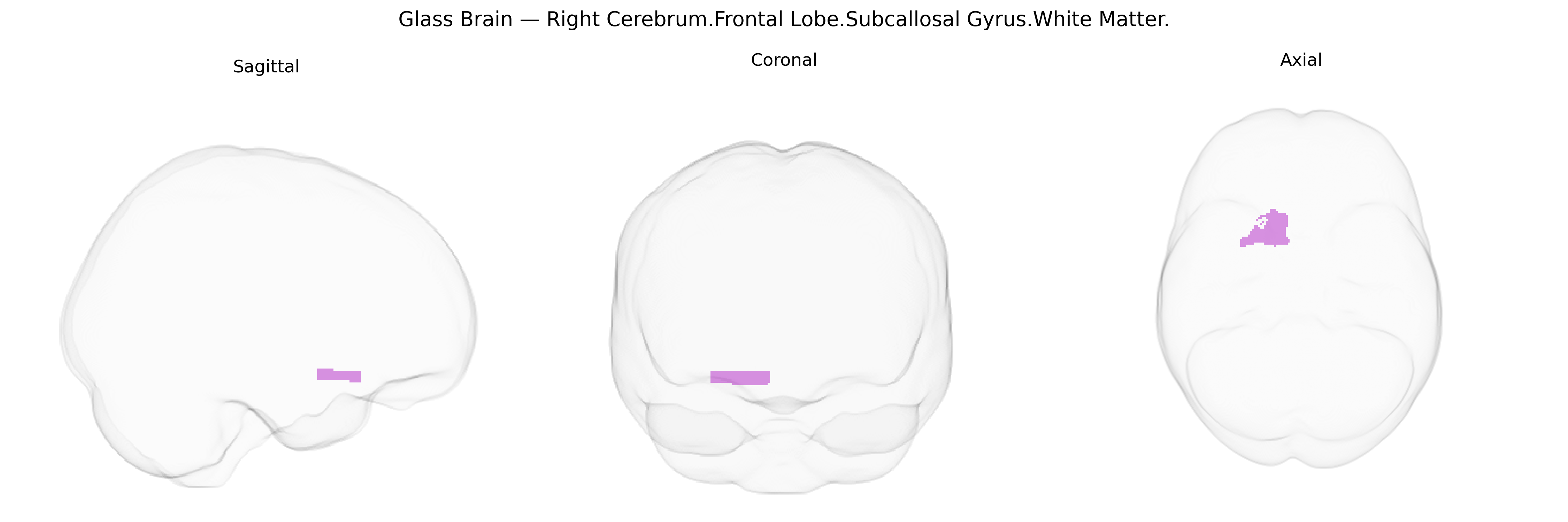
The bilateral Right Cerebrum.Frontal Lobe.Subcallosal Gyrus.White Matter corresponds to the deep myelinated fiber pathways underlying the subcallosal gyrus, a ventromedial frontal region located inferior to the genu of the corpus callosum and adjacent to the rostral anterior cingulate and orbitofrontal cortices. This white matter compartment contains association and projection fibers that interconnect limbic and prefrontal networks involved in emotional regulation, autonomic control, and reward-related processing, including pathways linking the medial prefrontal cortex, anterior cingulate, basal forebrain, and hypothalamic structures. There is no direct link for this specific white matter region; a closely related structure is the Subcallosal gyrus.
The Right Cerebrum Frontal Lobe Subcallosal Gyrus White Matter region, corresponding broadly to subgenual/ventromedial prefrontal and adjacent cingulo-frontal white matter pathways, has been repeatedly implicated in genetic studies of mood and affective disorders as well as structural brain GWAS. Large-scale imaging-genetics consortia (e.g., ENIGMA, UK Biobank) have reported SNP-based heritability for prefrontal white matter volume and integrity measures in this area, with associations involving genes related to myelination, axonal guidance, and synaptic plasticity (such as variants in CNTNAP2, NRG1, and genes within major histocompatibility and neurodevelopmental pathways), though specific loci for the exact Talairach-defined subcallosal white matter parcel are rarely isolated. GWAS of major depressive disorder, bipolar disorder, and neuroticism have implicated loci that also show effects on subgenual/ventromedial prefrontal thickness and white matter microstructure, suggesting shared genetic underpinnings between psychiatric risk and structural variation in this region. Polygenic risk scores for depression, bipolar disorder, and schizophrenia have been associated with reduced white matter integrity in frontal and cingulate tracts traversing the subcallosal area, such as parts of the cingulum bundle and frontostriatal pathways. Additionally, variants in serotonin-related and stress-response genes (e.g., SLC6A4, CRHR1) have been linked through candidate-gene and imaging-genetics studies to altered structure or connectivity in subgenual and subcallosal frontal regions, consistent with the well-established role of this circuitry in mood regulation, though these candidate associations have been less consistently replicated than more recent GWAS-based findings.
Overview generated by GPT-4o (2026).
Region ID: 291
Hemisphere: bilateral
Atlas: Talairach labels 1mm
Full Quality Version: Download MP4
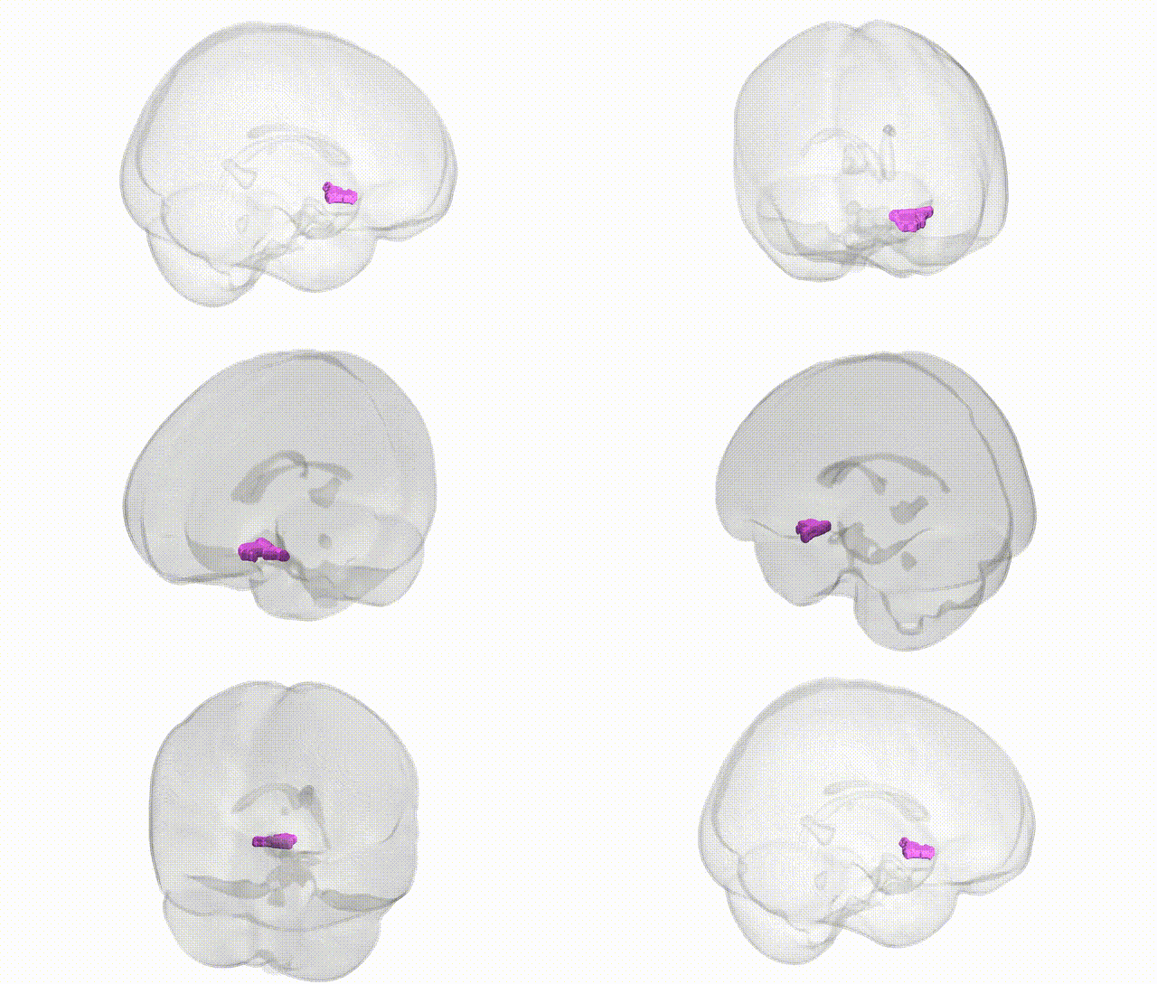
Full Quality Version: Download MP4
Wali Sidiqyar*, Gaurav Rudravaram*, Elyssa M. McMaster, Trent M. Schwartz, Adam M. Saunders, Kurt G. Schilling, Bennett A. Landman "Introducing SPINS: A Shared Public Visualization Library of Neuroanatomical Structures." Medical Imaging with Deep Learning- short paper
This resource is licensed under CC0 1.0 Universal (Public Domain).