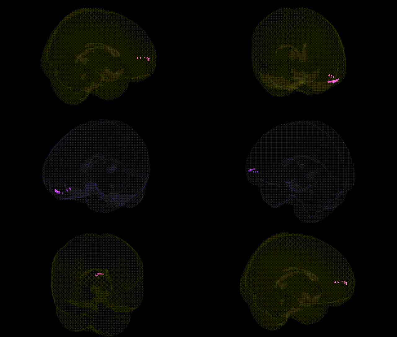
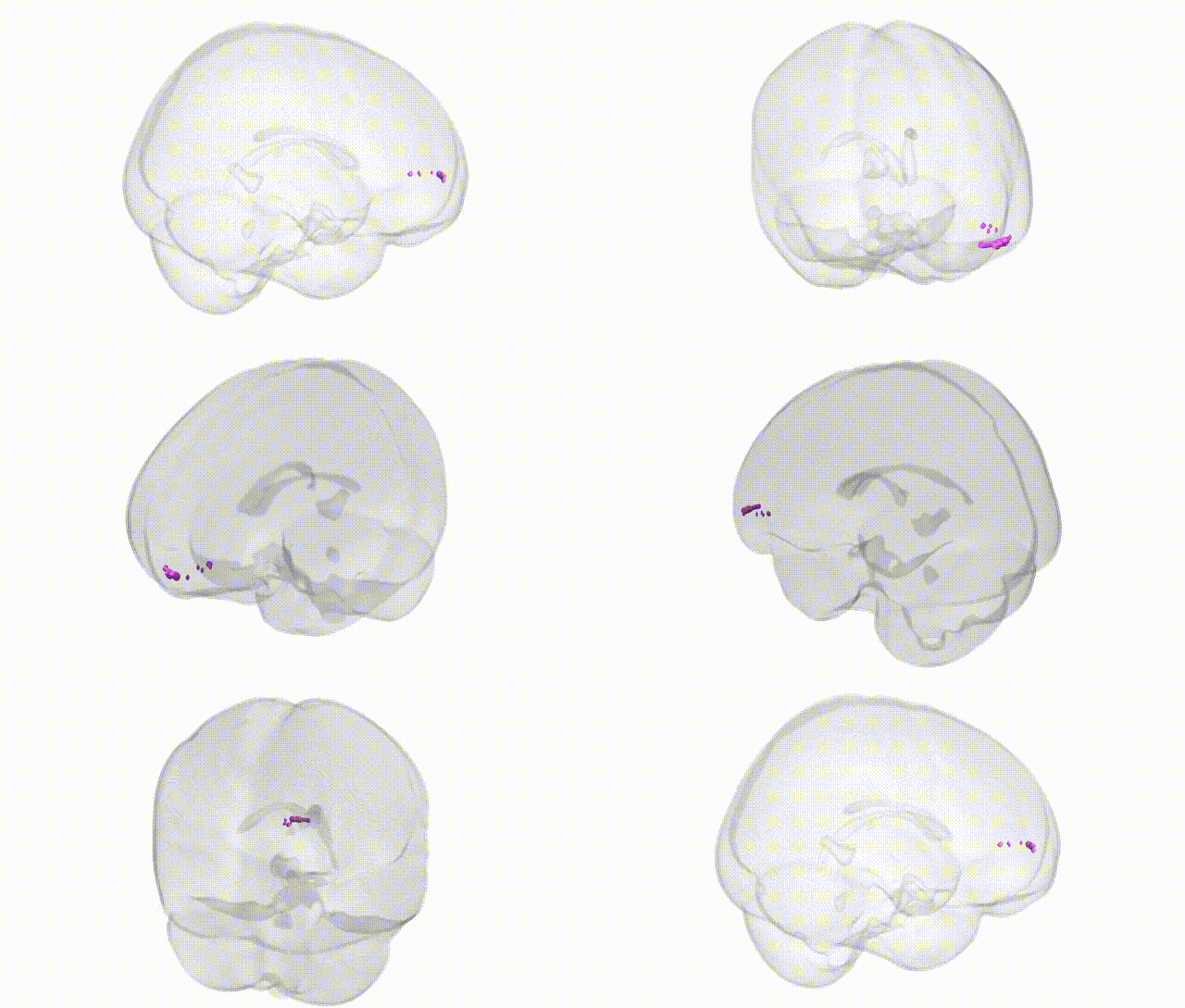
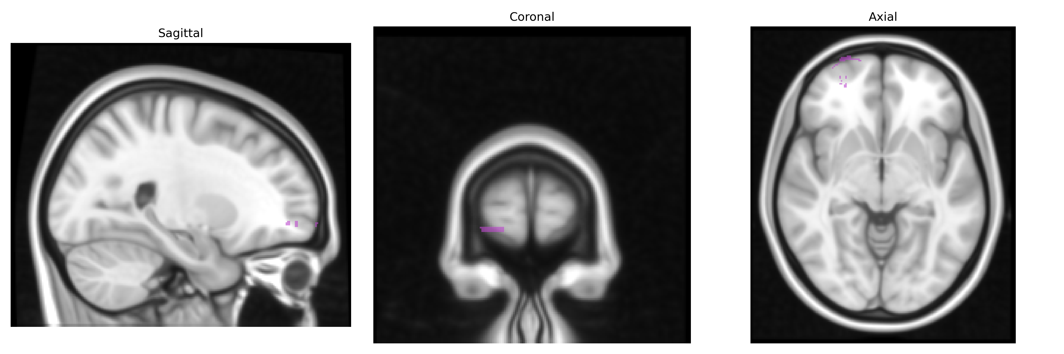
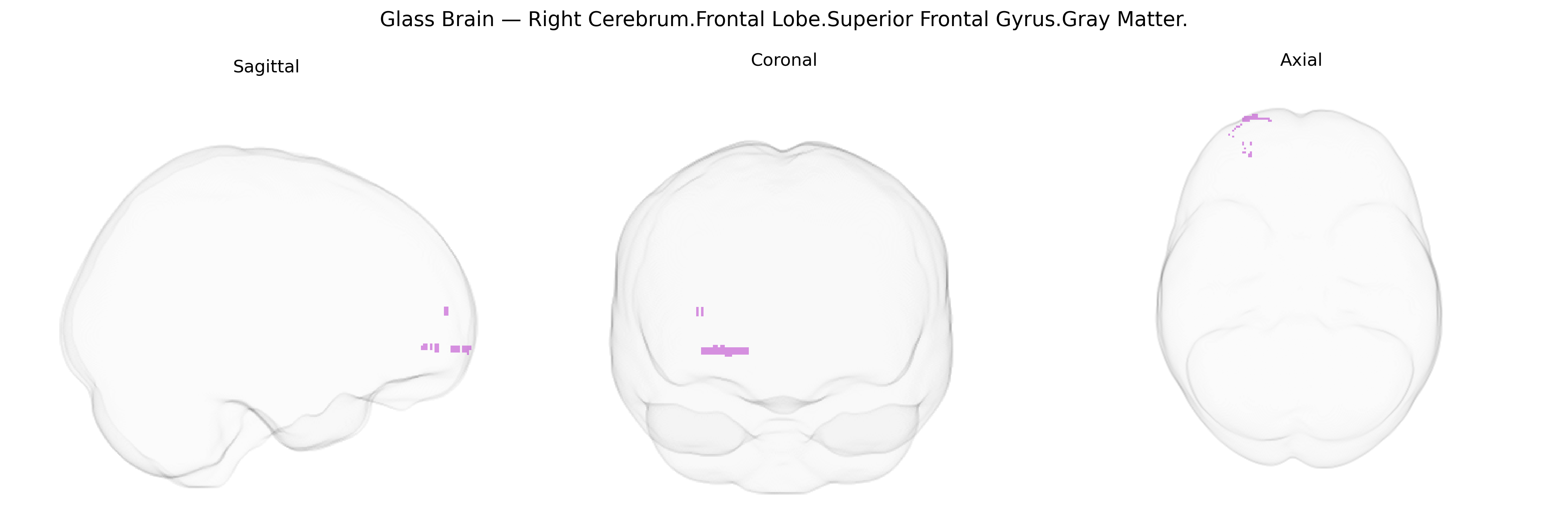
The bilateral Right Cerebrum.Frontal Lobe.Superior Frontal Gyrus.Gray Matter corresponds to the cortical gray matter of the superior frontal gyrus within the frontal lobe, extending along the dorsal aspect of the frontal cortex anterior to the premotor and supplementary motor areas. This region participates in higher-order executive functions, including working memory, attentional control, decision-making, and aspects of self-initiated behavior, and is also implicated in motor planning and the control of complex, goal-directed actions. Its anterior portions are associated with cognitive and social–emotional processes, while more posterior portions interface with premotor networks, reflecting its integrative role in linking cognition with action. Superior frontal gyrus
The bilateral Right Cerebrum.Frontal Lobe.Superior Frontal Gyrus gray matter region, as defined in the Talairach 1 mm atlas, has been implicated in multiple genetic studies of brain structure and function, although most work treats the superior frontal gyrus bilaterally or within broader frontal lobe or dorsomedial prefrontal cortex parcels rather than this exact label. Large GWAS of cortical morphology (e.g., ENIGMA, UK Biobank) have identified common variants in genes involved in neurodevelopment (such as HMGA2, IGF1, MAPT, and multiple loci near regulatory elements for synaptic and growth pathways) associated with superior frontal cortical thickness, surface area, and volume, which in turn show genetic correlations with general cognitive ability, educational attainment, and neuroticism. Polygenic risk for schizophrenia, bipolar disorder, major depression, ADHD, and autism spectrum disorder has been associated with altered superior frontal gray matter volume and cortical thickness in imaging–genetics studies, suggesting that part of the genetic liability for these disorders acts through this region’s structural and functional variation. Variants in genes affecting glutamatergic signaling (e.g., GRM3), calcium channel function (e.g., CACNA1C), and synaptic plasticity (e.g., BDNF) have been associated with prefrontal activation patterns and superior frontal involvement in executive control and working memory tasks. In Alzheimer’s disease and other dementias, GWAS risk loci such as APOE and BIN1 are associated with atrophy patterns that include superior frontal regions, while imaging–genetics work links these alleles to early cortical thinning and network disruption in frontal hubs. Across traits, the superior frontal gyrus emerges as a convergence point where heritable variation influencing intelligence, mood and anxiety phenotypes, risk-taking, and social cognition is reflected in genetically driven differences in gray matter structure and task-based or resting-state activation.
Overview generated by GPT-4o (2026).
Region ID: 613
Hemisphere: bilateral
Atlas: Talairach labels 1mm
Full Quality Version: Download MP4
Full Quality Version: Download MP4
Wali Sidiqyar*, Gaurav Rudravaram*, Elyssa M. McMaster, Trent M. Schwartz, Adam M. Saunders, Kurt G. Schilling, Bennett A. Landman "Introducing SPINS: A Shared Public Visualization Library of Neuroanatomical Structures." Medical Imaging with Deep Learning- short paper
This resource is licensed under CC0 1.0 Universal (Public Domain).