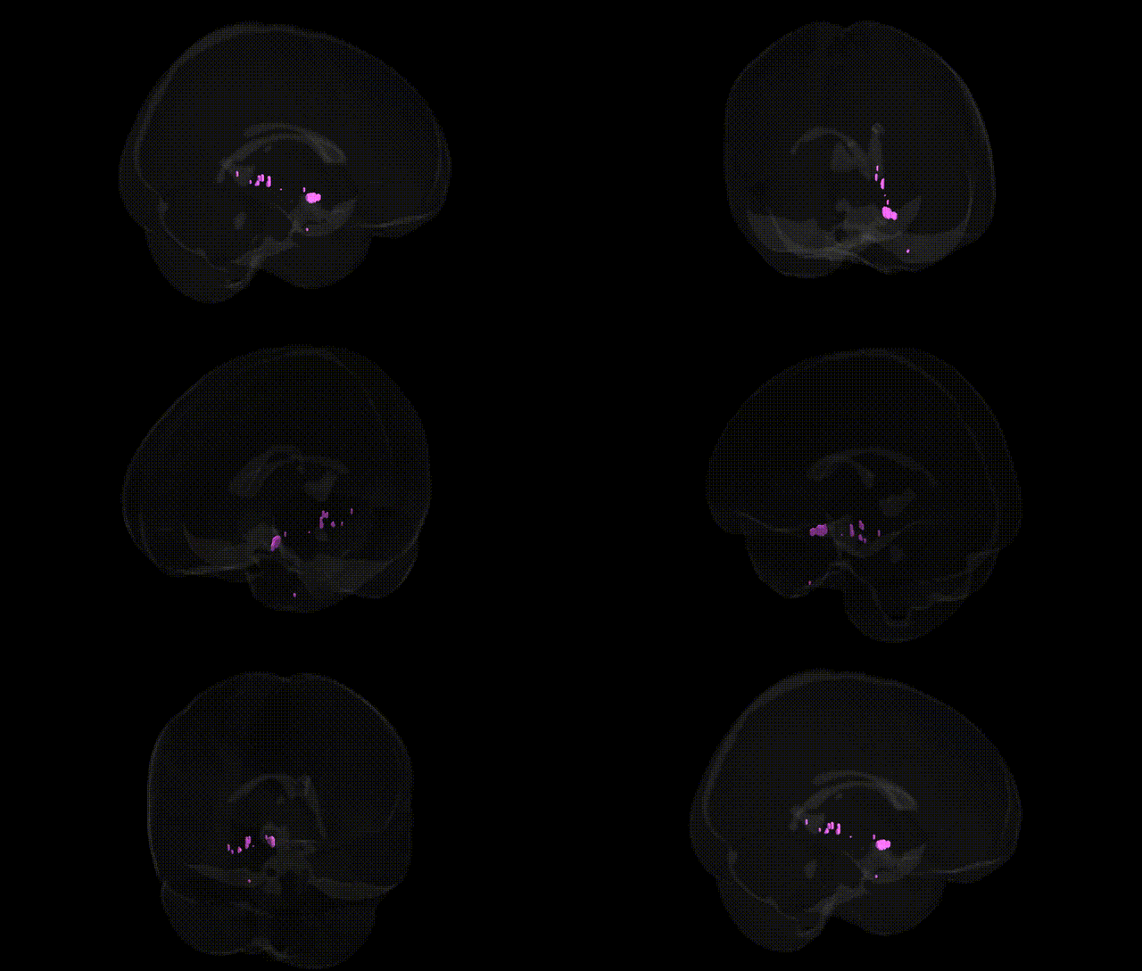
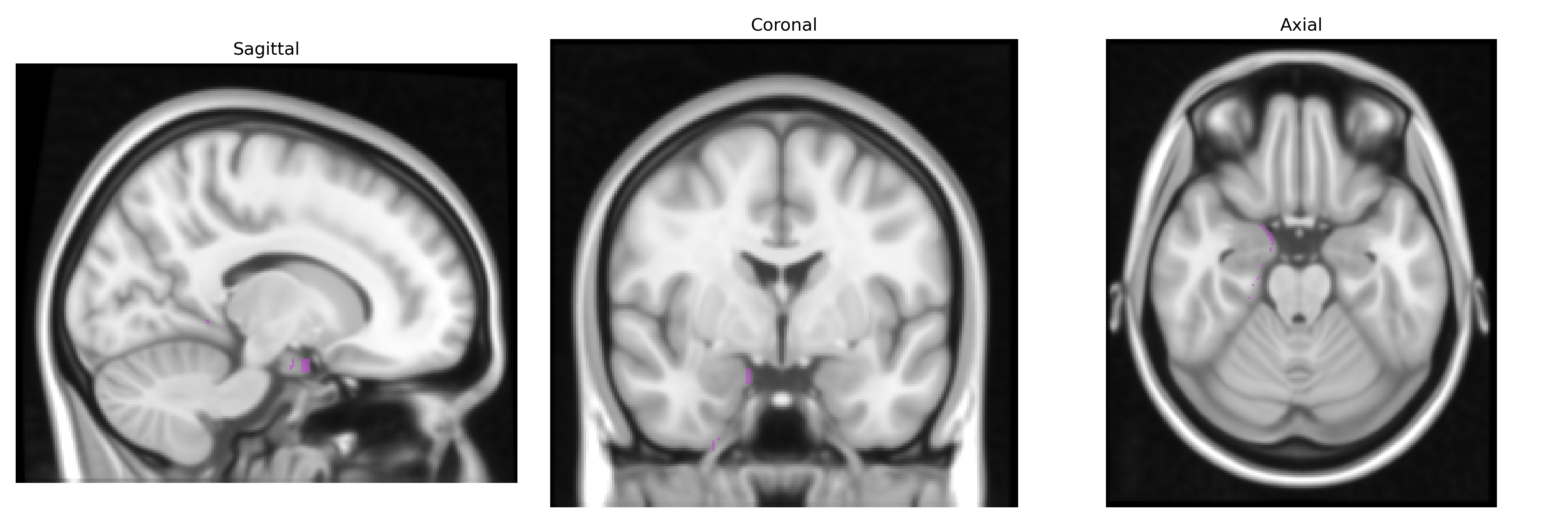
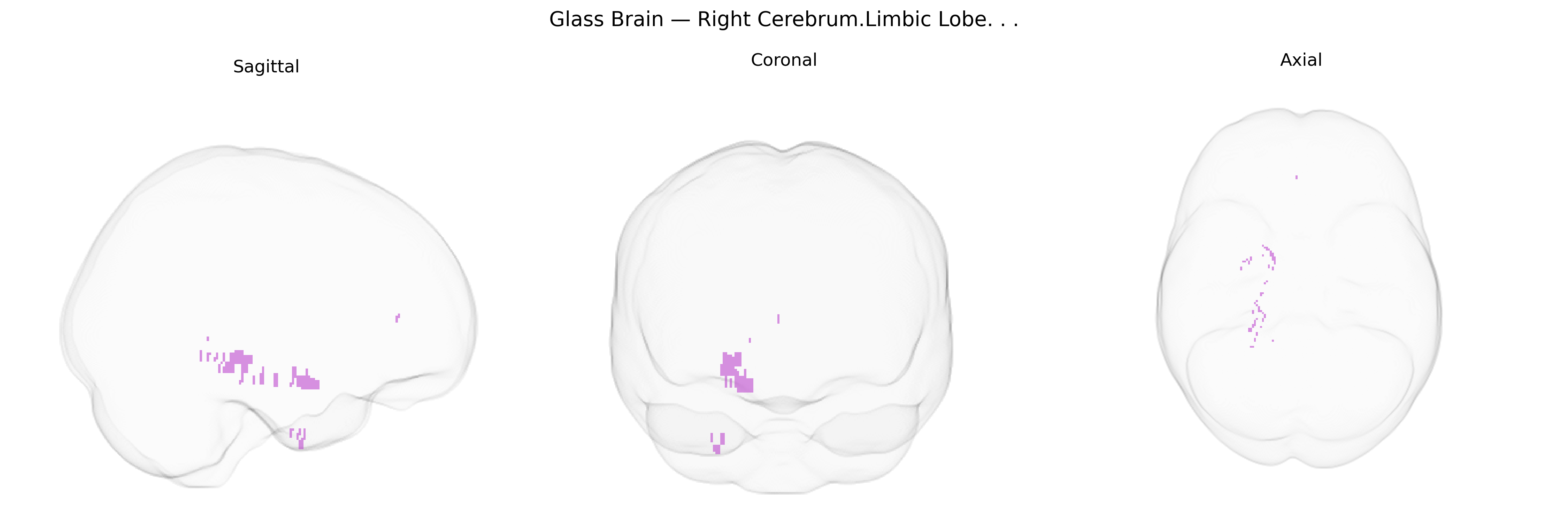
The bilateral Right Cerebrum.Limbic Lobe, as defined in the Talairach 1 mm atlas, refers to limbic lobe structures located in the right cerebral hemisphere, including portions of the cingulate gyrus, parahippocampal gyrus, and related medial temporal and medial frontal regions involved in emotion, memory, and autonomic regulation. Functionally, this territory participates in affective processing, encoding and retrieval of episodic memory, visceromotor integration, and aspects of motivation and behavior, often via strong connections with the hippocampus, amygdala, thalamus, and prefrontal cortex. These regions contribute to the limbic circuitry that integrates internal bodily states with cognitive and emotional context, supporting behavioral adaptation and emotional salience attribution. There is no direct link for this exact Talairach label; a related article is Limbic system.
The bilateral right cerebrum limbic lobe (as defined by the Talairach 1 mm atlas, encompassing structures such as the amygdala, hippocampus, parahippocampal gyrus, cingulate cortex, and related medial temporal regions) has been repeatedly implicated in imaging–genetics and GWAS-informed neuroimaging studies linking genetic variation to emotional, memory, and affective circuitry. Large consortia such as ENIGMA and UK Biobank have shown that common variants in and around genes like BDNF, APOE, SLC6A4, CACNA1C, and genes involved in glutamatergic and GABAergic signaling, synaptic plasticity, and neurodevelopment influence limbic volume, connectivity, and activity, particularly in the hippocampus and amygdala. These limbic traits show significant SNP-heritability and overlap genetically with risk for major depressive disorder, bipolar disorder, schizophrenia, post-traumatic stress disorder, anxiety disorders, Alzheimer’s disease, and other dementias, as well as personality traits such as neuroticism and harm avoidance. Amygdala- and hippocampus-focused GWAS have identified loci (for example in genes such as WWOX, MSRB3, HRK, and others) associated with structural differences in these regions, while polygenic risk scores for depression, schizophrenia, and Alzheimer’s disease correlate with altered limbic morphology and functional reactivity. Although most GWAS target specific limbic structures rather than the entire “right limbic lobe” label, convergent evidence supports a broad genetic architecture in which multiple common variants, often of small effect, influence right-sided limbic anatomy and function and, through this circuitry, confer susceptibility to a range of psychiatric and neurodegenerative disorders.
Overview generated by GPT-4o (2026).
Region ID: 22
Hemisphere: bilateral
Atlas: Talairach labels 1mm
Full Quality Version: Download MP4
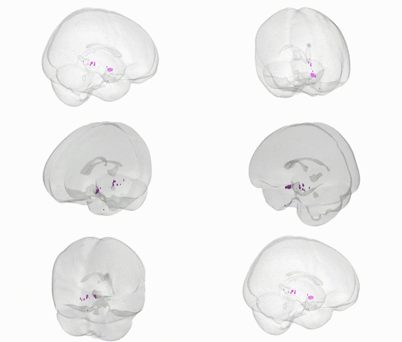
Full Quality Version: Download MP4
Wali Sidiqyar*, Gaurav Rudravaram*, Elyssa M. McMaster, Trent M. Schwartz, Adam M. Saunders, Kurt G. Schilling, Bennett A. Landman "Introducing SPINS: A Shared Public Visualization Library of Neuroanatomical Structures." Medical Imaging with Deep Learning- short paper
This resource is licensed under CC0 1.0 Universal (Public Domain).