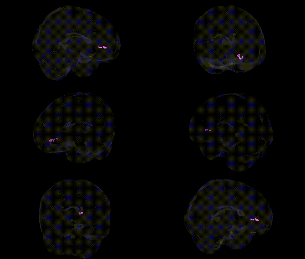
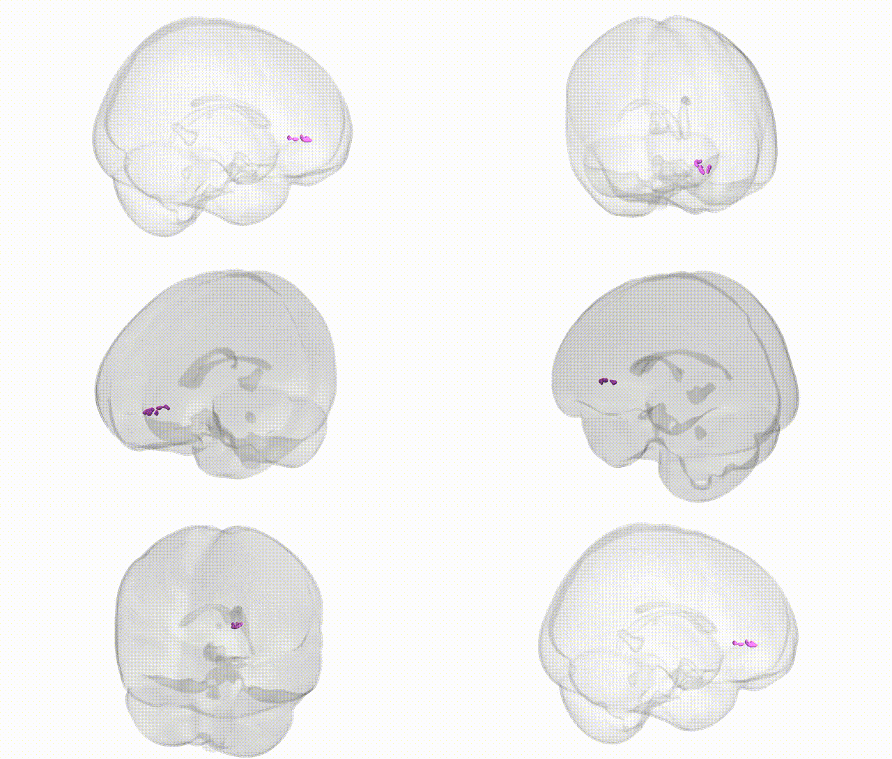
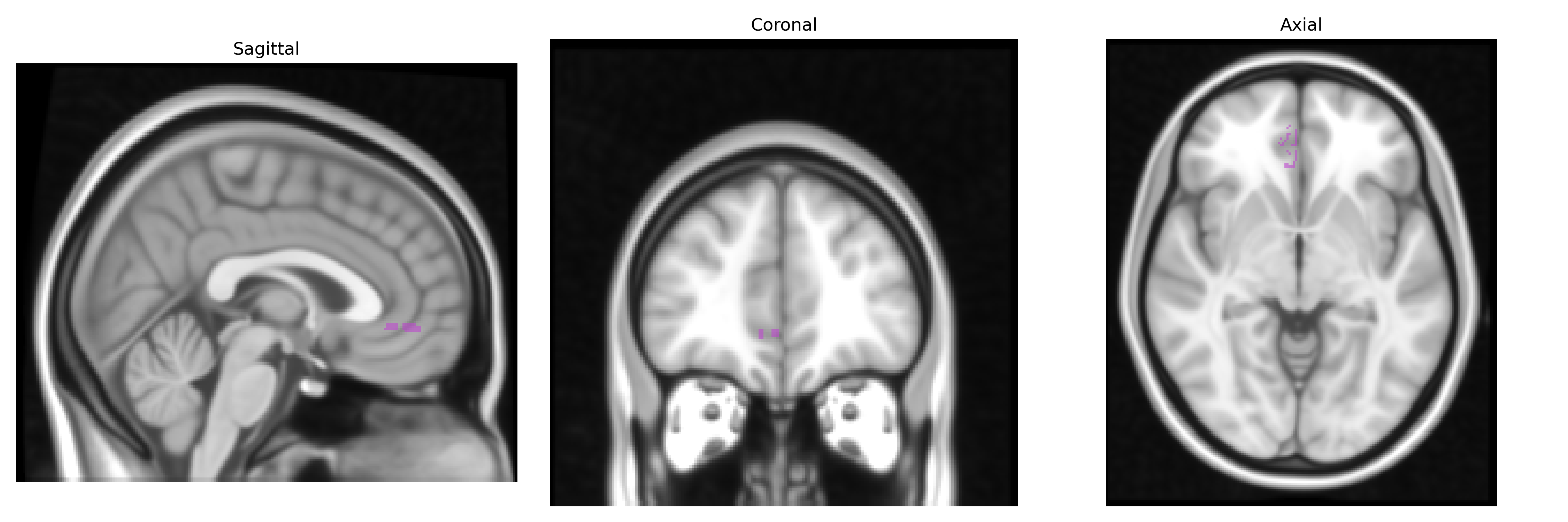
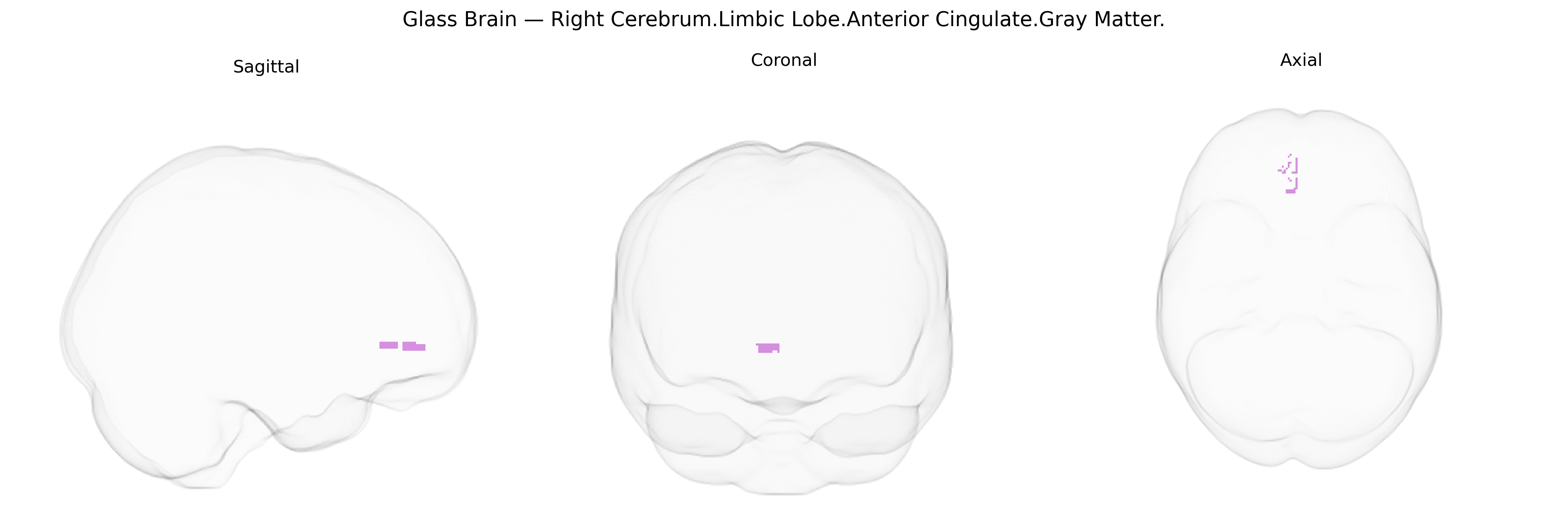
The bilateral Right Cerebrum.Limbic Lobe.Anterior Cingulate.Gray Matter, as labeled in the Talairach 1 mm atlas, corresponds to gray matter within the anterior portion of the cingulate cortex located on the medial aspect of the frontal lobe, arching over the corpus callosum. This region is a key node of the limbic system and is involved in affective processing, autonomic regulation, error detection, conflict monitoring, and motivational aspects of behavior, including the integration of emotional and cognitive information for decision-making. Functionally, it participates in attentional control, pain perception (particularly the affective dimension of pain), and regulation of emotional responses, in part through extensive connectivity with prefrontal, limbic, and subcortical structures such as the amygdala and basal ganglia. There is no direct link for this exact Talairach label; a closely related structure is the Anterior cingulate cortex.
The bilateral right anterior cingulate gray matter (Talairach: Right Cerebrum, Limbic Lobe, Anterior Cingulate) has been repeatedly implicated in imaging-genetics and GWAS-based neuroanatomical studies, though most findings refer to cingulate or anterior cingulate cortex more generally rather than this exact atlas label. Variants in genes related to synaptic plasticity, glutamatergic and GABAergic signaling, and neurodevelopment (for example, CACNA1C, GRM3, NRG1, and genes in the major histocompatibility complex region) have been associated with volumetric and cortical thickness variation in anterior cingulate regions, and with structural and functional connectivity involving this area. Large-scale ENIGMA consortium analyses and other GWAS of cortical morphology have identified multiple loci (e.g., near genes such as HMGA2, MIR548F3, and others involved in neuronal growth and patterning) that contribute to interindividual differences in cingulate thickness and surface area, though these loci are not specific to the right anterior cingulate alone. Genetically influenced alterations of this region’s structure and function are reported across a range of disorders, including major depressive disorder, bipolar disorder, schizophrenia, autism spectrum disorder, and attention-deficit/hyperactivity disorder, where risk loci (e.g., in CACNA1C, ZNF804A, DRD2, SLC6A4, BDNF, and polygenic risk scores for these conditions) correlate with anterior cingulate gray matter volume, activity, or connectivity. Additionally, genetic risk for chronic pain, anxiety, and neuroticism, as well as for cognitive traits such as executive function and educational attainment, has been linked to variation in anterior cingulate structure and activation in GWAS and polygenic score studies, underscoring this region’s role as a genetically modulated hub for affective regulation, cognitive control, and pain processing rather than a site with a single disorder- or gene-specific association.
Overview generated by GPT-4o (2026).
Region ID: 606
Hemisphere: bilateral
Atlas: Talairach labels 1mm
Full Quality Version: Download MP4
Full Quality Version: Download MP4
Wali Sidiqyar*, Gaurav Rudravaram*, Elyssa M. McMaster, Trent M. Schwartz, Adam M. Saunders, Kurt G. Schilling, Bennett A. Landman "Introducing SPINS: A Shared Public Visualization Library of Neuroanatomical Structures." Medical Imaging with Deep Learning- short paper
This resource is licensed under CC0 1.0 Universal (Public Domain).