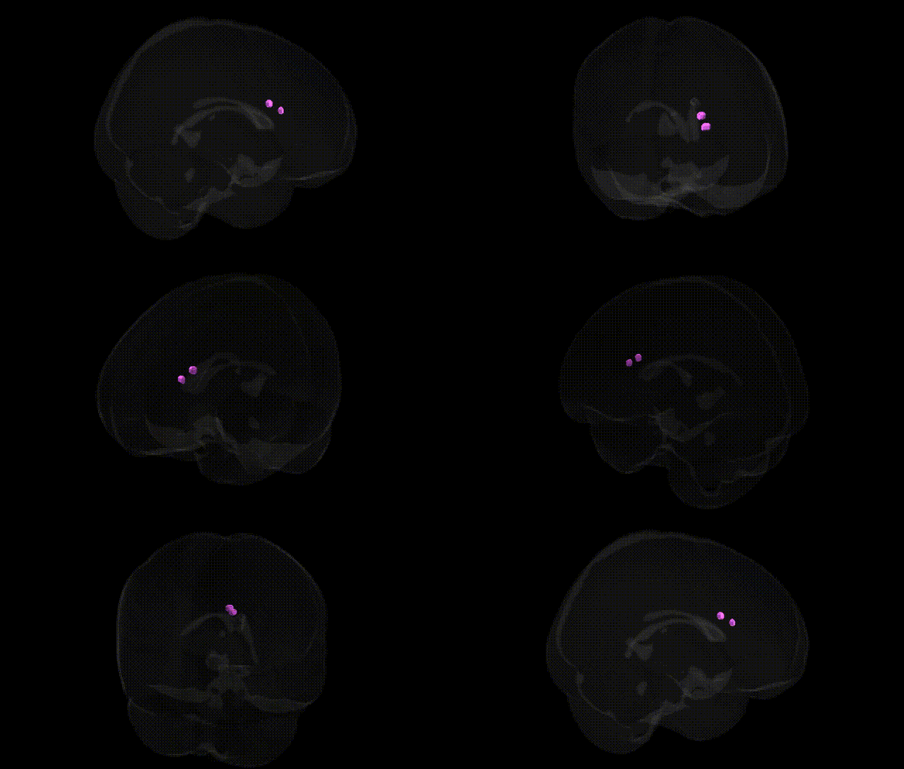
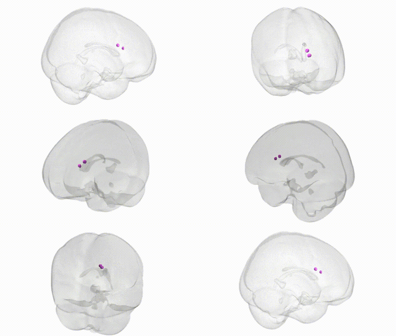
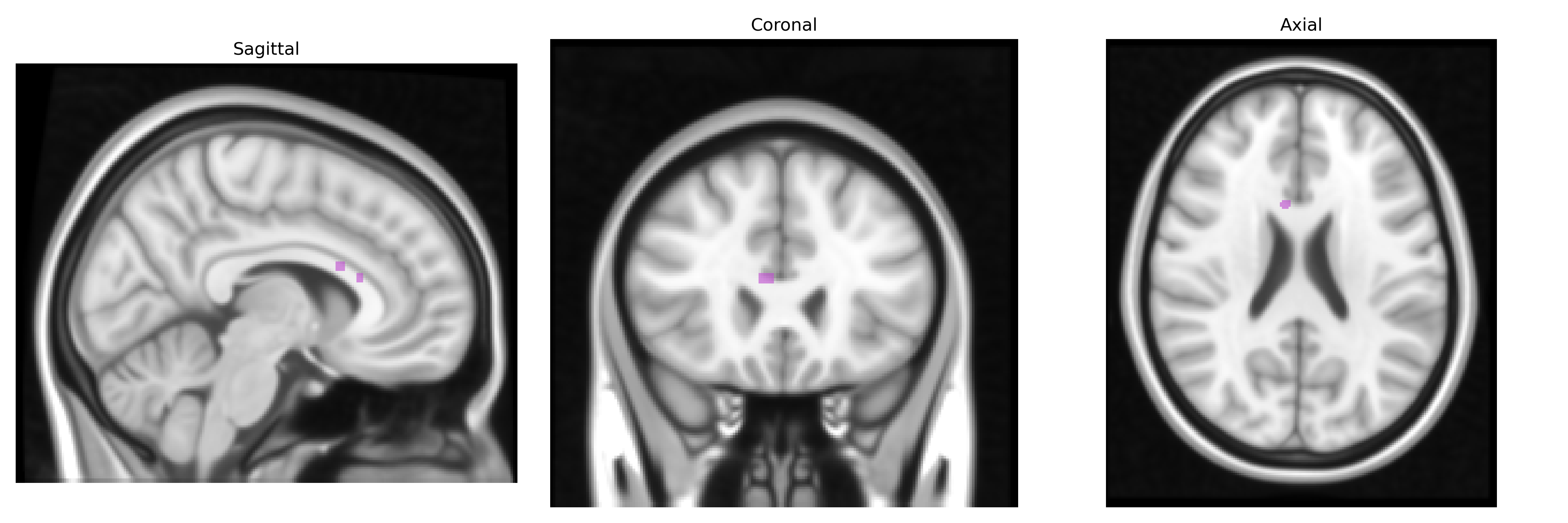
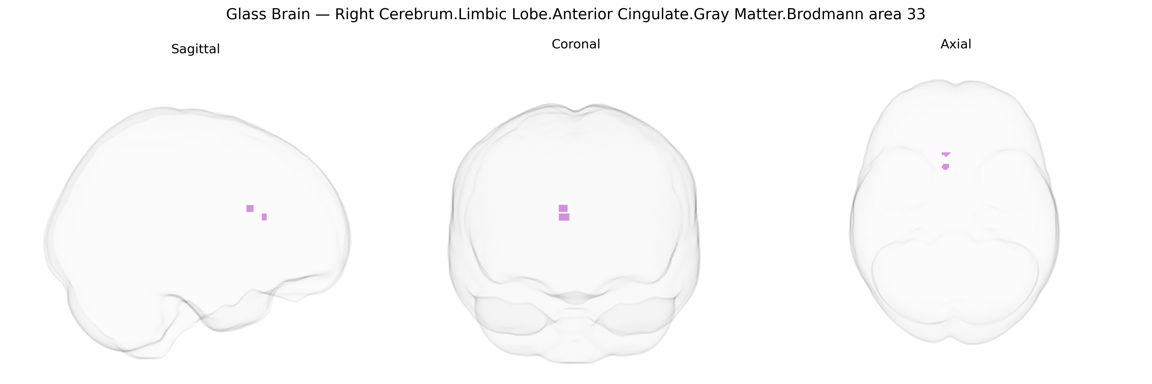
The bilateral Right Cerebrum.Limbic Lobe.Anterior Cingulate.Gray Matter.Brodmann area 33 corresponds to a small cytoarchitectonic field located in the ventral portion of the anterior cingulate gyrus, adjacent to the genu of the corpus callosum and embedded within the limbic lobe. It is considered part of the ventral anterior cingulate cortex and is implicated in affective and autonomic components of pain processing, emotional evaluation of stimuli, and modulation of visceromotor responses. Neuronal populations in this region integrate limbic input from structures such as the amygdala and medial prefrontal cortex and project to autonomic and basal forebrain systems, contributing to regulation of emotional behavior, stress responses, and aspects of internal state monitoring. There is no direct link for Brodmann area 33; a related structure is the Anterior cingulate cortex.
The bilateral Right Cerebrum Limbic Lobe Anterior Cingulate Gray Matter Brodmann area 33, a subregion of the anterior cingulate cortex (ACC), has been implicated in genetic associations largely through studies targeting the broader ACC or medial prefrontal/limbic networks rather than BA33 specifically; nonetheless, convergent evidence links common variants in serotonin (e.g., 5-HTTLPR in SLC6A4), dopamine (e.g., COMT Val158Met), and glutamatergic genes (e.g., GRIN2B) to structural and functional variation in this region, including gray matter volume and task-evoked activity related to emotion, pain, and conflict processing. Large-scale GWAS and imaging-genetics consortia (e.g., ENIGMA, UK Biobank) have identified polygenic influences on ACC thickness and volume, with notable contributions from loci near genes involved in neurodevelopment, synaptic plasticity, and myelination, and these ACC-related genetic profiles overlap partially with polygenic risk for major depressive disorder, schizophrenia, bipolar disorder, anxiety traits, and chronic pain conditions. ACC—encompassing BA33—has repeatedly emerged in GWAS-informed imaging studies of depression and negative affect (e.g., associations with polygenic scores for depression and neuroticism predicting reduced ACC volume or altered connectivity), and in candidate-gene and polygenic analyses of pain sensitivity and placebo response, where genetic variation in opioid (OPRM1) and catecholamine-related genes moderates ACC activity during nociception. Additionally, schizophrenia and psychosis risk alleles (e.g., in CACNA1C, ZNF804A, and other GWAS-implicated loci) show associations with altered ACC structure or functional coupling within cingulo-frontal networks, which include BA33, suggesting that genetic risk for these disorders partly manifests through development and function of this limbic ACC territory.
Overview generated by GPT-4o (2026).
Region ID: 839
Hemisphere: bilateral
Atlas: Talairach labels 1mm
Full Quality Version: Download MP4
Full Quality Version: Download MP4
Wali Sidiqyar*, Gaurav Rudravaram*, Elyssa M. McMaster, Trent M. Schwartz, Adam M. Saunders, Kurt G. Schilling, Bennett A. Landman "Introducing SPINS: A Shared Public Visualization Library of Neuroanatomical Structures." Medical Imaging with Deep Learning- short paper
This resource is licensed under CC0 1.0 Universal (Public Domain).