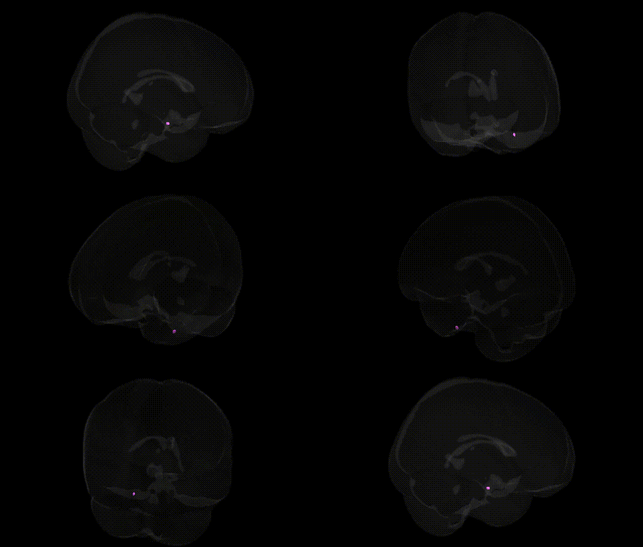
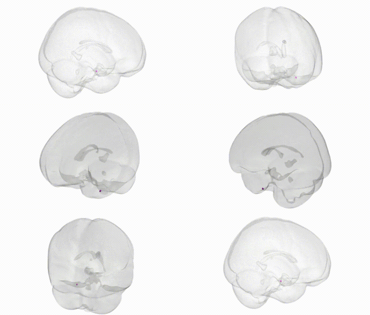
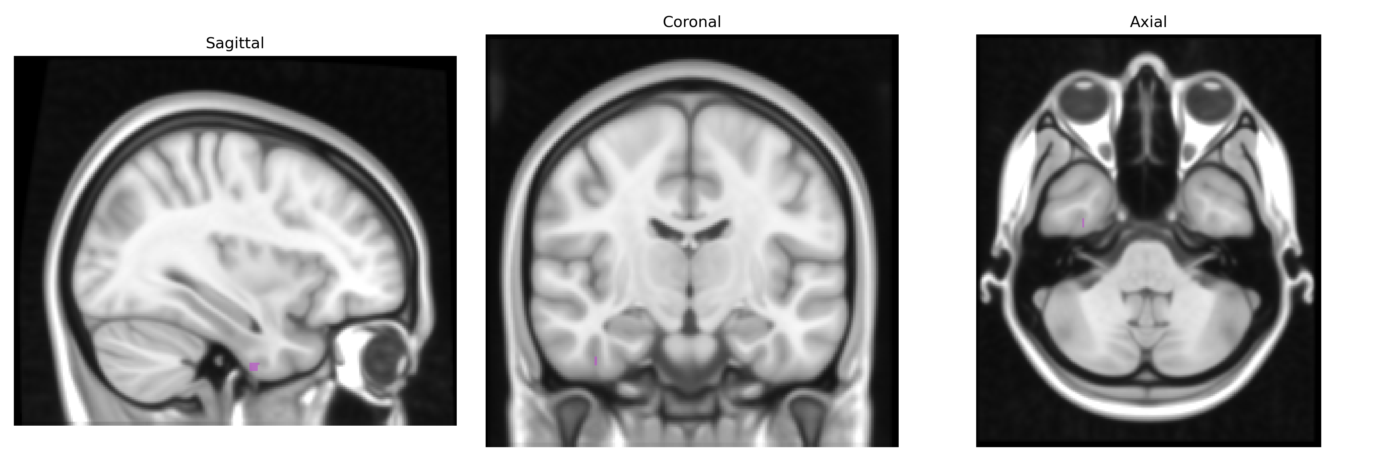
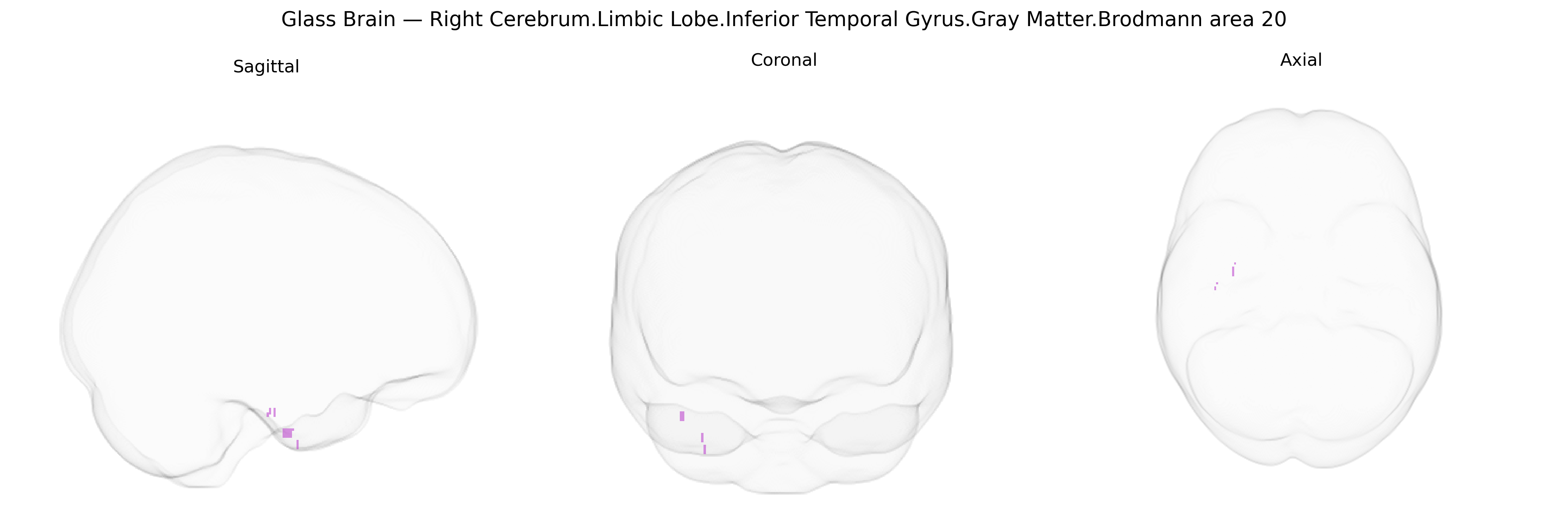
The bilateral Right Cerebrum.Limbic Lobe.Inferior Temporal Gyrus.Gray Matter.Brodmann area 20 corresponds to a portion of the ventral temporal cortex involved in higher-order visual and associative processing, including object recognition, semantic integration, and aspects of visual memory. Situated on the inferior surface of the temporal lobe, this cortex lies anterior and lateral to primary visual areas and receives multimodal input from visual, auditory, and limbic structures, supporting the linkage of complex visual stimuli with stored knowledge and emotional significance. Neuronal populations in this region exhibit selectivity for complex features and contribute to category-level representations (such as faces and objects), while extensive reciprocal connectivity with hippocampal and parahippocampal regions facilitates consolidation and retrieval of episodic and semantic information. There is no direct link; see the related area Brodmann area 20.
The bilateral right inferior temporal gyrus of the limbic/ventral temporal region, encompassing Brodmann area 20, has emerged in imaging–genetics and GWAS-based neuroimaging studies as a locus where common variants influence cortical thickness, surface area, and gray-matter volume, often in the context of broader temporal and limbic networks rather than this parcel alone. Large MRI–GWAS consortia (e.g., ENIGMA, UK Biobank) have implicated loci in or near genes involved in neurodevelopment (such as those in synaptic formation, axon guidance, and neuronal migration pathways) that show significant associations with temporal lobe morphology, including inferior temporal gyri, with some of these loci overlapping risk architecture for schizophrenia, major depressive disorder, and bipolar disorder. Polygenic risk scores for Alzheimer’s disease and other dementias have been associated with structural variation in temporal lobe regions including BA20, consistent with the region’s role in object recognition and semantic memory, while autism spectrum disorder and dyslexia–related genetic risk has also been linked to alterations in ventral temporal and inferior temporal cortices in imaging–genetic studies. In addition, variants affecting genes like APOE, CLU, and others involved in lipid metabolism and amyloid processing, although not specific to BA20, show downstream structural and functional effects on temporal and limbic cortices that encompass this area. Overall, genetic findings point to BA20 as part of a distributed, polygenic network in which risk alleles for neurodevelopmental, psychiatric, and neurodegenerative disorders modulate gray matter structure and connectivity rather than acting on this region in isolation.
Overview generated by GPT-4o (2026).
Region ID: 19
Hemisphere: bilateral
Atlas: Talairach labels 1mm
Full Quality Version: Download MP4
Full Quality Version: Download MP4
Wali Sidiqyar*, Gaurav Rudravaram*, Elyssa M. McMaster, Trent M. Schwartz, Adam M. Saunders, Kurt G. Schilling, Bennett A. Landman "Introducing SPINS: A Shared Public Visualization Library of Neuroanatomical Structures." Medical Imaging with Deep Learning- short paper
This resource is licensed under CC0 1.0 Universal (Public Domain).