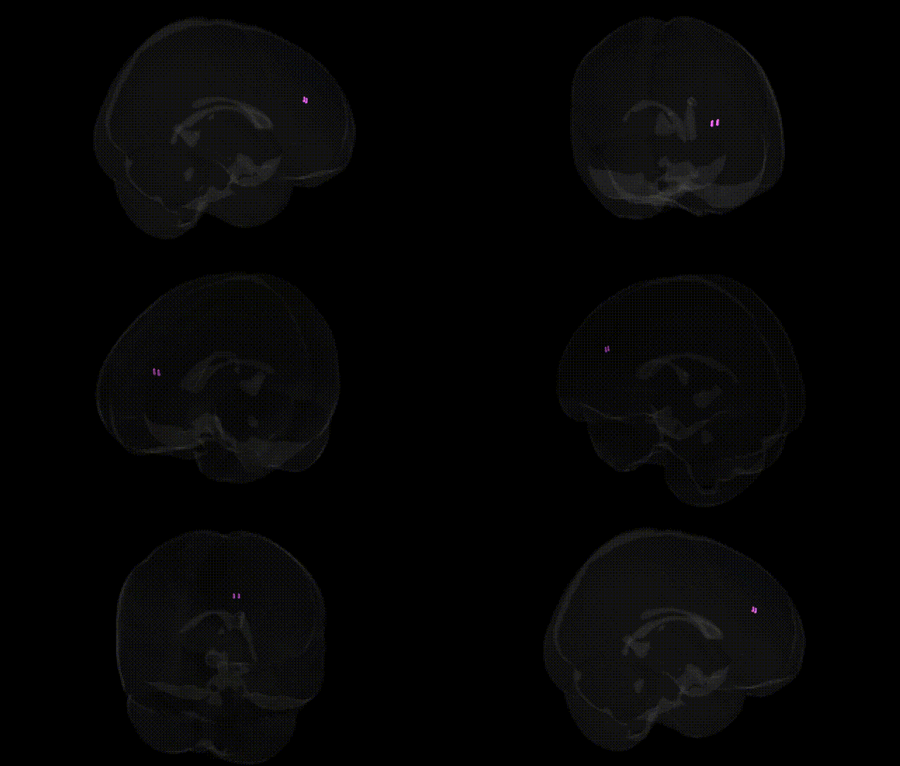
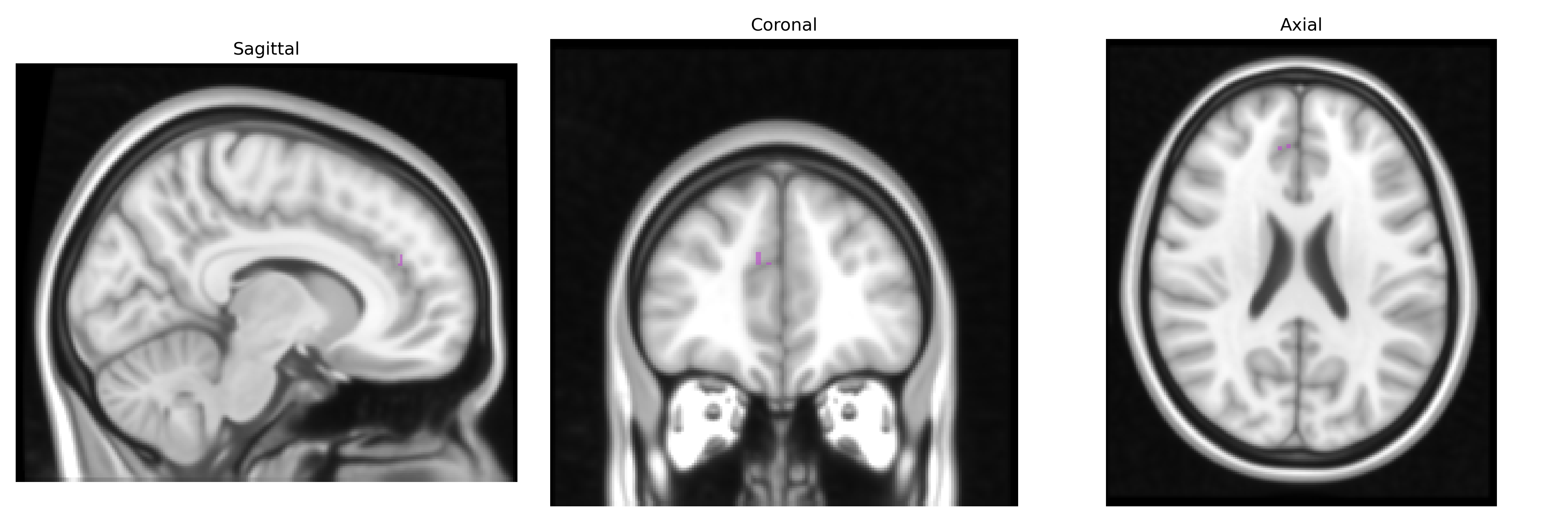
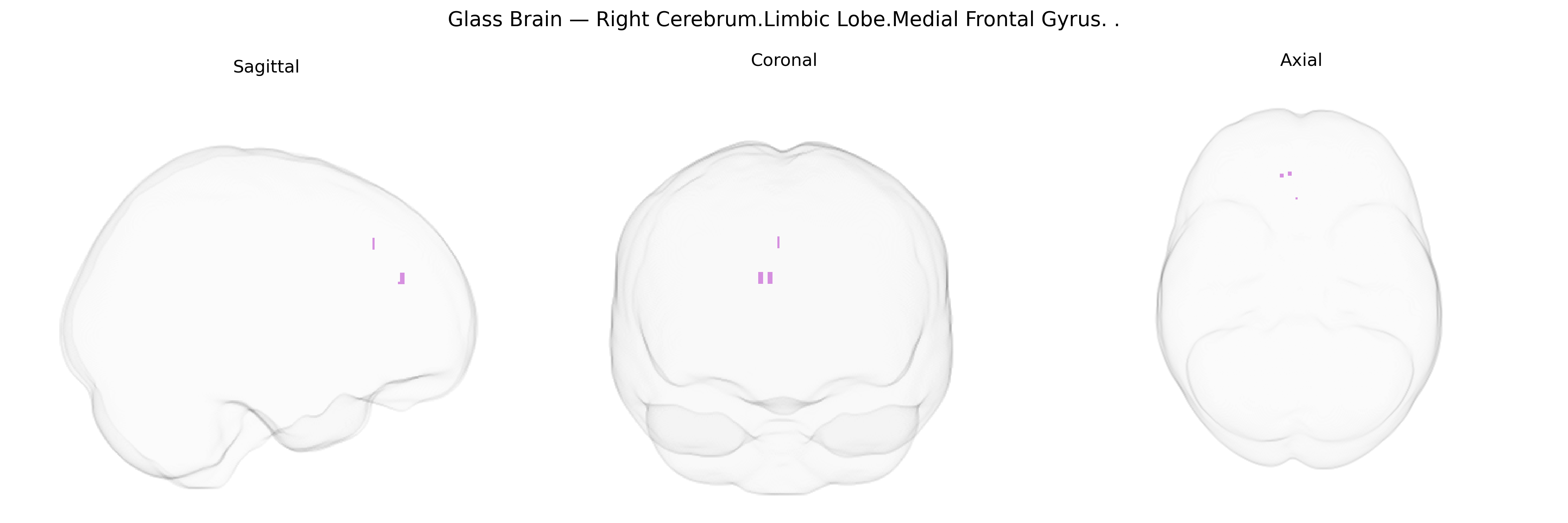
The bilateral Right Cerebrum.Limbic Lobe.Medial Frontal Gyrus corresponds largely to the medial portion of the frontal cortex on the right side, encompassing parts of the medial prefrontal and anterior cingulate regions that are functionally associated with the limbic system. This area participates in higher-order emotional processing, valuation of actions and outcomes, motivation, and aspects of self-referential and social cognition, often integrating affective information with executive control. It is also implicated in regulation of autonomic responses, decision-making under uncertainty, and modulation of stress and mood states, with extensive reciprocal connections to limbic structures such as the amygdala, hippocampus, and other cingulate territories. There is no direct link for “Medial Frontal Gyrus”; a related structure is the Medial prefrontal cortex.
The bilateral medial frontal gyrus of the limbic lobe (right cerebrum), as defined in the Talairach labels 1 mm atlas, has been implicated in multiple genetic and genome-wide association studies via its roles in medial prefrontal, anterior cingulate, and ventromedial prefrontal networks involved in emotion regulation, decision-making, and self-referential processing. Polygenic risk for major depressive disorder, schizophrenia, bipolar disorder, and anxiety-related traits repeatedly associates with altered structure or function in this region, with significant contributions from genes related to synaptic plasticity and glutamatergic transmission (e.g., CACNA1C, GRM3, NRG1, and other schizophrenia/bipolar risk loci) and serotonergic and stress-response pathways (e.g., SLC6A4, HTR1A, FKBP5) that modulate medial prefrontal reactivity to emotional and stress-related stimuli. Large neuroimaging–genetics consortia (such as ENIGMA and UK Biobank) have shown that common variants influencing cortical thickness and surface area in medial frontal regions overlap with risk loci for psychiatric disorders and cognitive traits, and GWAS of resting-state networks identify associations between medial prefrontal connectivity and polygenic scores for depression, neuroticism, and subjective well-being. Additionally, medial frontal morphometry and activation patterns modulated by APOE and other Alzheimer’s disease risk variants link this region to early neurodegenerative changes, while dopaminergic and frontostriatal genes (e.g., COMT, DRD2/DRD4) have been connected to medial frontal activation during tasks requiring cognitive control, reward processing, and error monitoring.
Overview generated by GPT-4o (2026).
Region ID: 950
Hemisphere: bilateral
Atlas: Talairach labels 1mm
Full Quality Version: Download MP4
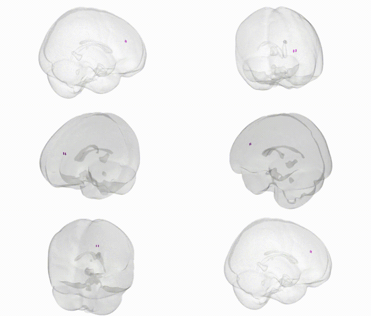
Full Quality Version: Download MP4
Wali Sidiqyar*, Gaurav Rudravaram*, Elyssa M. McMaster, Trent M. Schwartz, Adam M. Saunders, Kurt G. Schilling, Bennett A. Landman "Introducing SPINS: A Shared Public Visualization Library of Neuroanatomical Structures." Medical Imaging with Deep Learning- short paper
This resource is licensed under CC0 1.0 Universal (Public Domain).