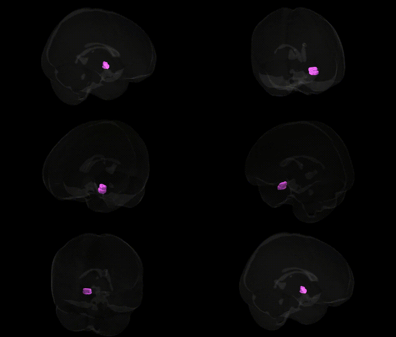
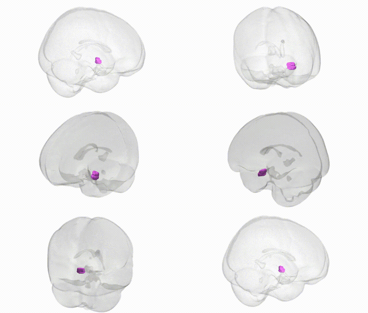
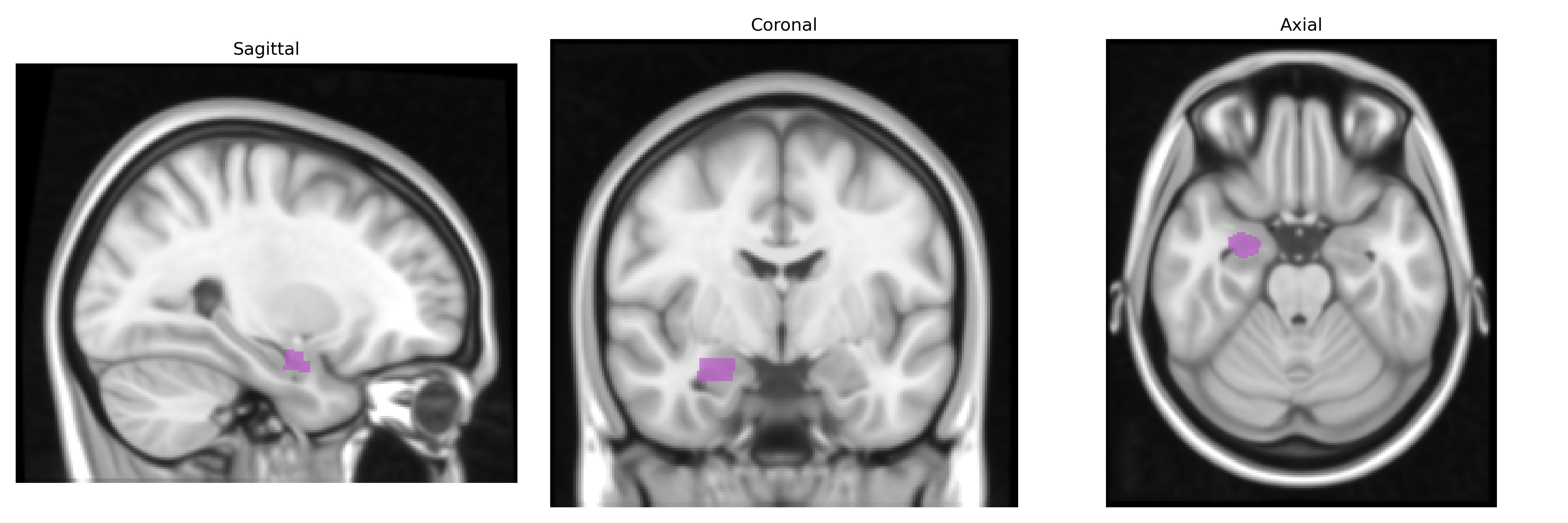
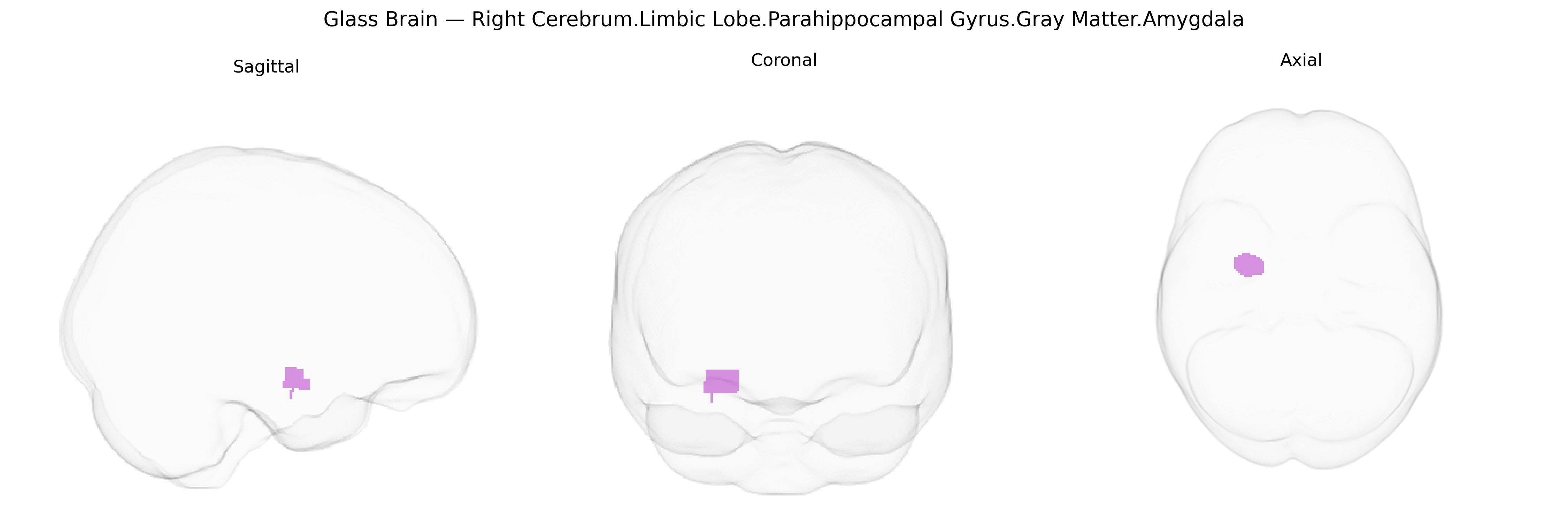
The bilateral Right Cerebrum.Limbic Lobe.Parahippocampal Gyrus.Gray Matter.Amygdala corresponds to the amygdaloid complex, a gray-matter structure situated deep within the medial temporal lobe and closely associated with the parahippocampal gyrus. It comprises multiple nuclei (including basolateral, centromedial, and cortical groups) that integrate sensory, visceral, and contextual information to modulate emotional processing, especially fear and threat detection, as well as associative learning and emotional memory. The amygdala has dense reciprocal connections with the hippocampus, prefrontal cortex, hypothalamus, and brainstem autonomic centers, enabling coordination of endocrine, autonomic, and behavioral responses to emotionally salient stimuli. In the Talairach 1 mm atlas, this label denotes the gray-matter component of the amygdala within the limbic lobe, encompassing both hemispheres despite the “Right Cerebrum” designation in the hierarchical path. Amygdala
The bilateral right parahippocampal gyrus and amygdala (Talairach 1 mm atlas limbic gray matter region) have been implicated in multiple genetic and GWAS findings, particularly in relation to psychiatric, neurodevelopmental, and neurodegenerative traits. Common variants in genes such as BDNF, SLC6A4, COMT, and 5-HT receptor genes, as well as polygenic risk for major depressive disorder, anxiety disorders, post-traumatic stress disorder, bipolar disorder, and schizophrenia, have been associated with altered amygdala and parahippocampal volume or reactivity in imaging–genetics studies. Large-scale GWAS of subcortical and limbic structures (e.g., ENIGMA consortium) have identified loci in or near genes involved in synaptic plasticity, neurodevelopment, and calcium signaling (such as DLG2, HMGA2, SLC39A8, and others) that correlate with amygdala and parahippocampal morphology, though many associations are shared across limbic and cortical regions rather than strictly specific to this parcel. Genetic risk for Alzheimer’s disease (including APOE ε4 and polygenic risk scores) and other dementias is linked to early atrophy and functional disruption in the parahippocampal–amygdala complex, while risk variants for autism spectrum disorder and social behavior traits have been associated with amygdala volume and connectivity differences. Overall, the region is a key structural and functional node where polygenic influences on emotion, memory, stress sensitivity, and neurodegeneration converge, but most identified loci exert modest, distributed effects rather than region-exclusive associations.
Overview generated by GPT-4o (2026).
Region ID: 183
Hemisphere: bilateral
Atlas: Talairach labels 1mm
Full Quality Version: Download MP4
Full Quality Version: Download MP4
Wali Sidiqyar*, Gaurav Rudravaram*, Elyssa M. McMaster, Trent M. Schwartz, Adam M. Saunders, Kurt G. Schilling, Bennett A. Landman "Introducing SPINS: A Shared Public Visualization Library of Neuroanatomical Structures." Medical Imaging with Deep Learning- short paper
This resource is licensed under CC0 1.0 Universal (Public Domain).