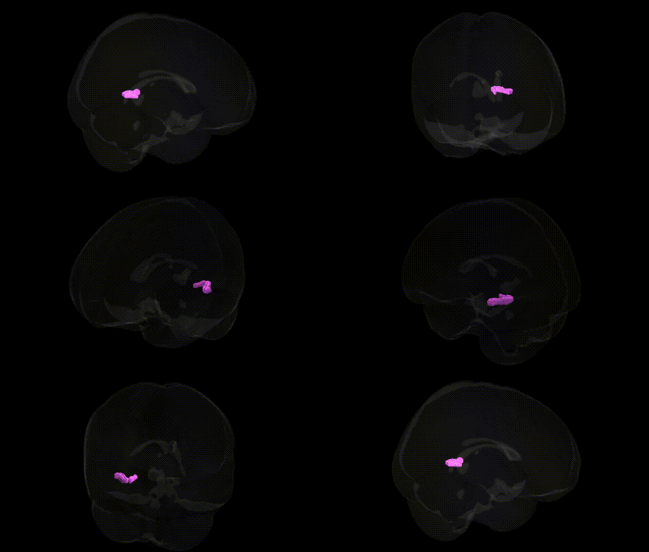
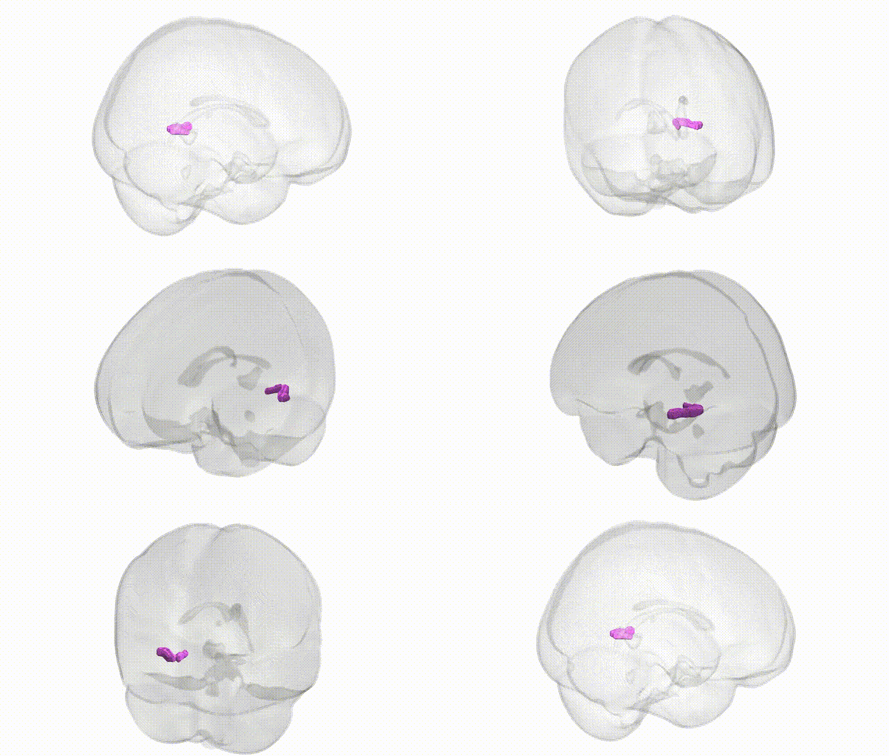
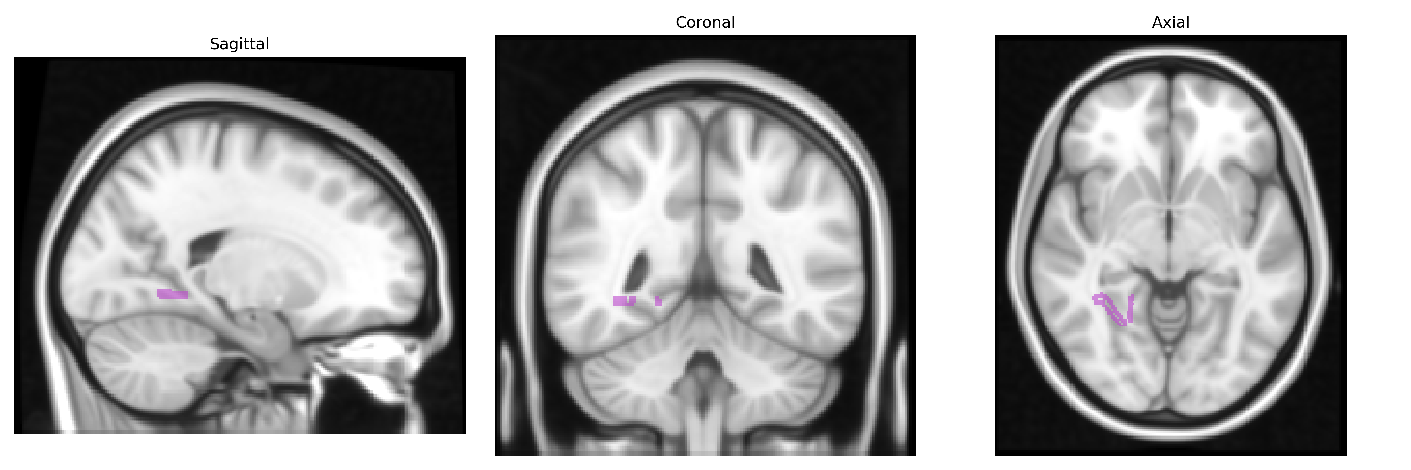
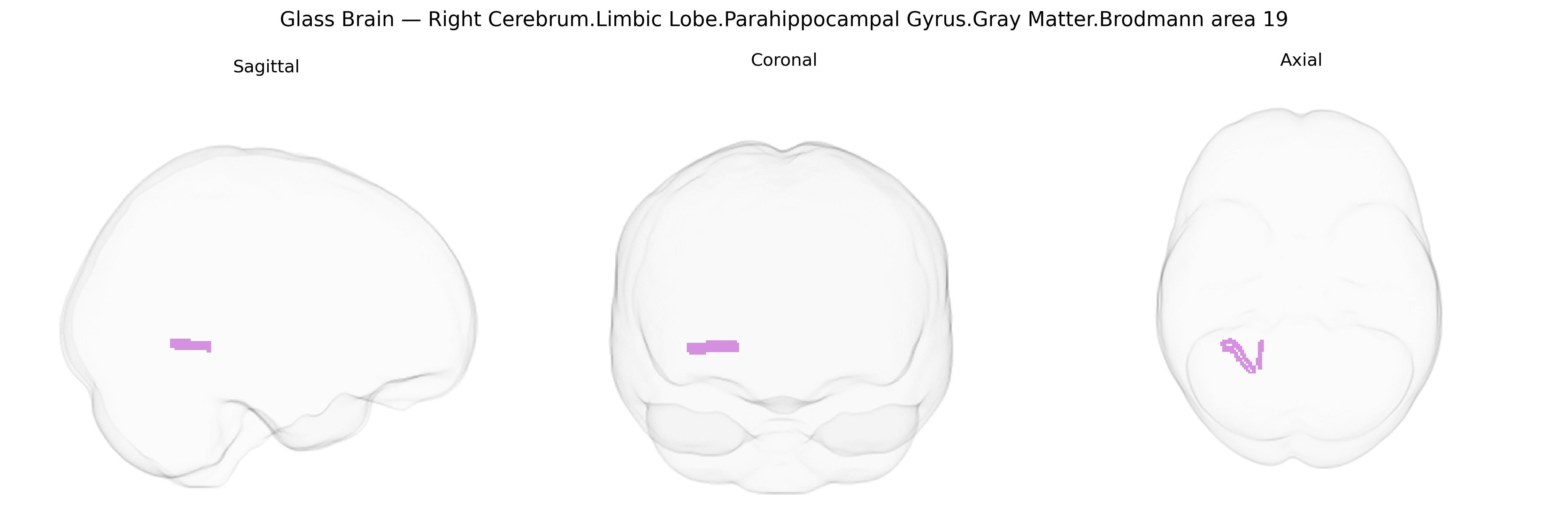
The bilateral Right Cerebrum.Limbic Lobe.Parahippocampal Gyrus.Gray Matter.Brodmann area 19 region, as defined in the Talairach 1 mm atlas, represents a gray-matter parcel located at the interface of the parahippocampal gyrus and occipitotemporal visual association cortex, reflecting a mapping extension of Brodmann area 19 into limbic/medial temporal territory. Functionally, this region is associated with higher-order visual processing, including complex scene analysis and visuospatial integration, while also interacting with limbic structures involved in contextual memory and navigation, such as the hippocampal formation. Its position within the parahippocampal gyrus suggests a role in linking visual context with episodic memory and environmental layout, consistent with its connectivity to both visual association areas and medial temporal memory circuits. There is no direct Wikipedia article for this exact composite region; a closely related structure is the Parahippocampal gyrus.
The bilateral right parahippocampal gyrus gray matter extending into Brodmann area 19, as defined in the Talairach 1 mm atlas, lies at the intersection of medial temporal and occipital cortices and has been implicated in genetic studies of memory, visuospatial processing, and neuropsychiatric risk via both region-of-interest and voxel-based morphometry GWAS. Variants in genes involved in synaptic plasticity and neurodevelopment (including BDNF, COMT, and APOE, as well as multiple loci identified in large ENIGMA and UK Biobank imaging-genetics consortia) show associations with parahippocampal and adjacent occipital/BA19 gray matter volume or cortical thickness, which in turn correlate with episodic memory performance and scene/face processing. Polygenic risk scores for Alzheimer’s disease, schizophrenia, major depressive disorder, and autism spectrum disorder have been linked to structural alterations in parahippocampal and nearby visual-association regions, suggesting shared genetic influences on limbic–visual integration circuits; Alzheimer’s-associated loci such as APOE ε4 and CLU are particularly tied to reduced medial temporal/parahippocampal integrity. GWAS of brain imaging phenotypes also highlight multiple common variants across the genome—often in regulatory regions near neurodevelopmental and synaptic genes—that contribute to interindividual differences in gray matter density and surface area in this region, and these structural measures mediate genetic effects on cognitive traits such as spatial navigation, memory encoding, and susceptibility to age-related cognitive decline.
Overview generated by GPT-4o (2026).
Region ID: 426
Hemisphere: bilateral
Atlas: Talairach labels 1mm
Full Quality Version: Download MP4
Full Quality Version: Download MP4
Wali Sidiqyar*, Gaurav Rudravaram*, Elyssa M. McMaster, Trent M. Schwartz, Adam M. Saunders, Kurt G. Schilling, Bennett A. Landman "Introducing SPINS: A Shared Public Visualization Library of Neuroanatomical Structures." Medical Imaging with Deep Learning- short paper
This resource is licensed under CC0 1.0 Universal (Public Domain).