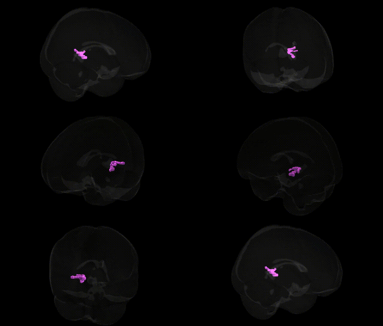
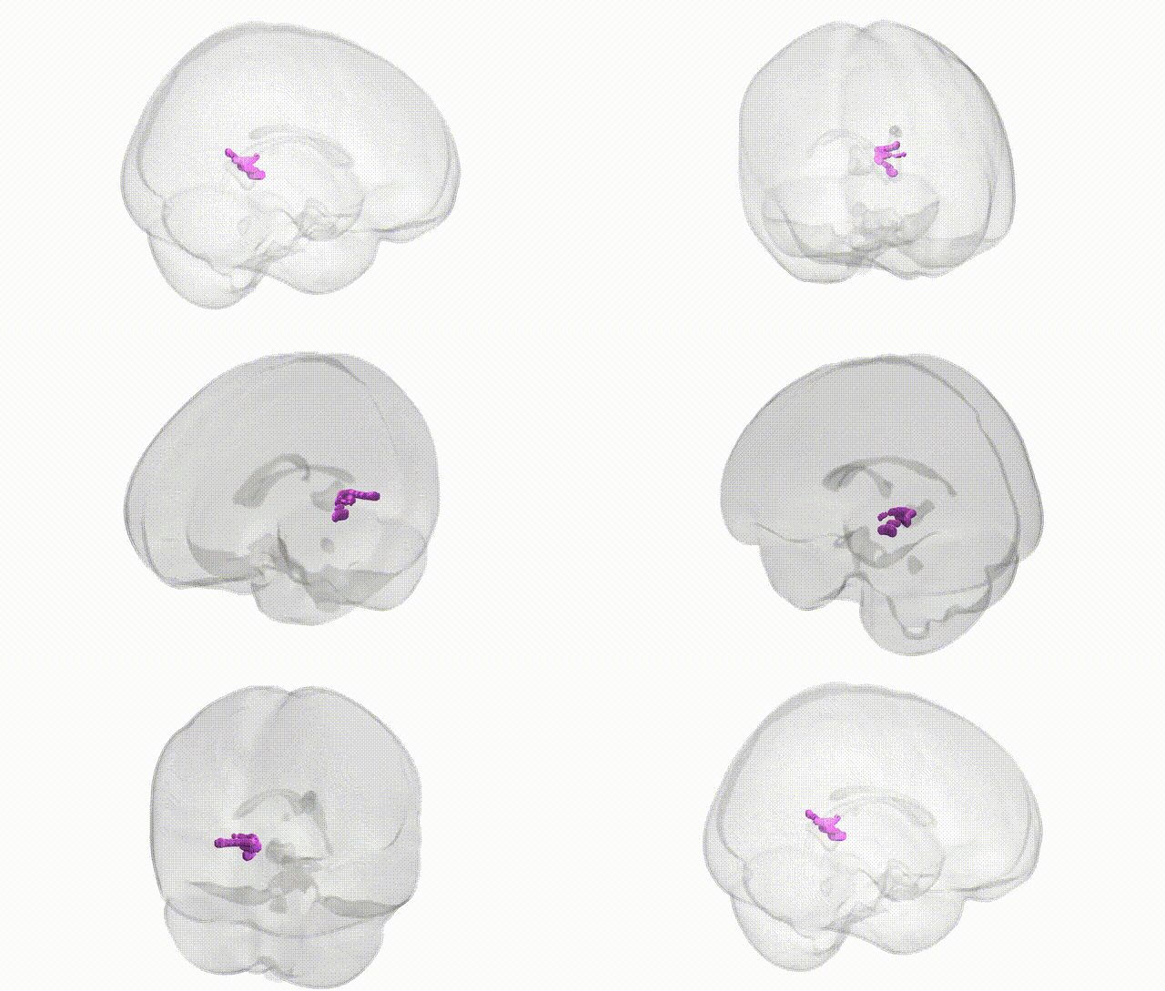
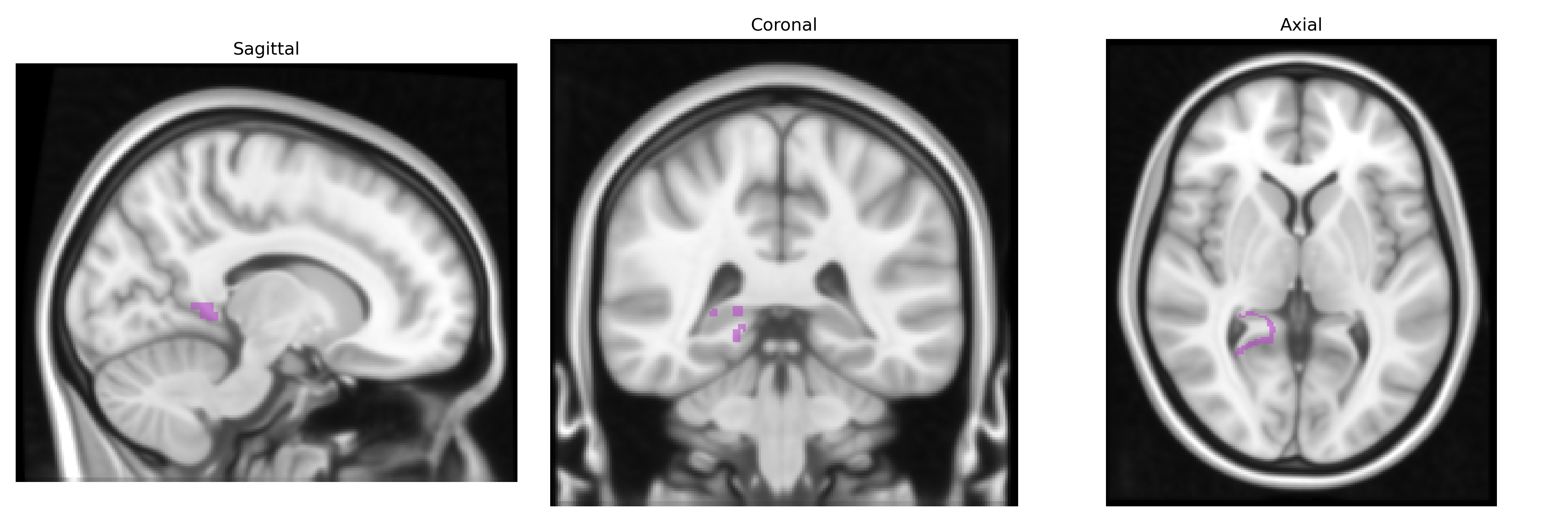
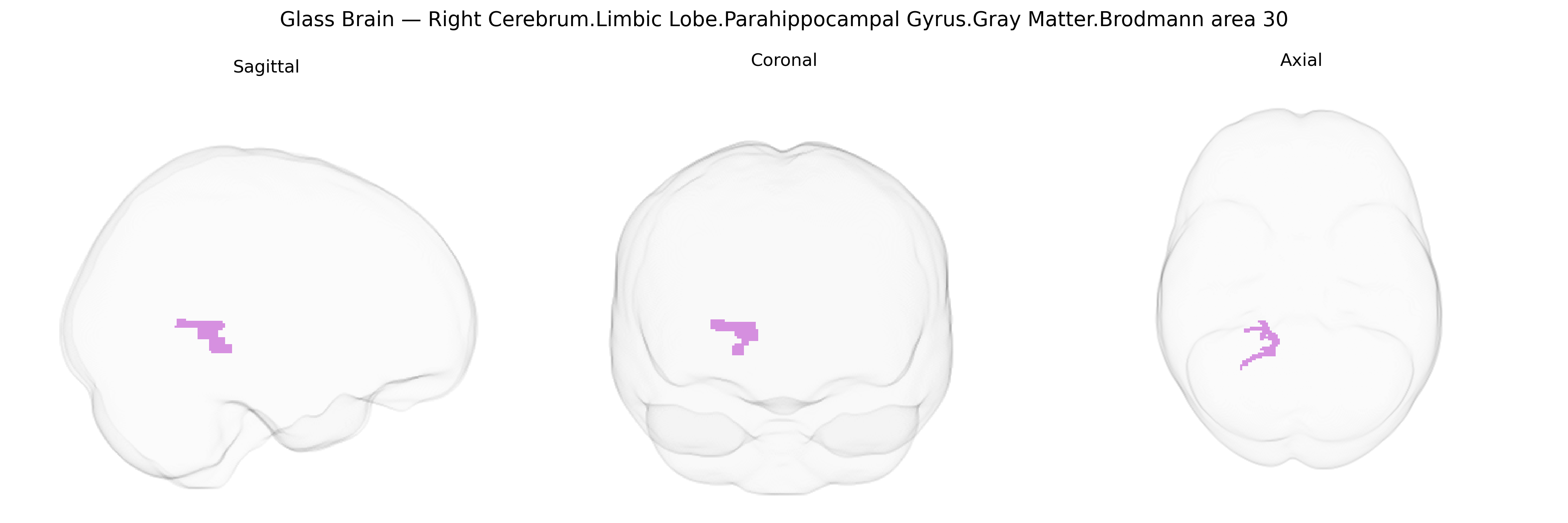
The bilateral Right Cerebrum.Limbic Lobe.Parahippocampal Gyrus.Gray Matter.Brodmann area 30 corresponds to the retrosplenial cortex portion of the parahippocampal gyrus, a limbic-associated cortical field located on the medial surface of the temporal–occipital junction, adjacent to the isthmus of the cingulate gyrus and bordering Brodmann areas 23 and 29. Histologically, BA30 is a granular or dysgranular cortex involved in integrating visuospatial, mnemonic, and contextual information, forming part of a broader retrosplenial–hippocampal–parahippocampal network critical for episodic memory, spatial navigation, and scene processing. Functionally, this region participates in encoding and retrieval of contextual associations, orientation within familiar environments, and coupling internal mnemonic representations with external visual cues, especially via reciprocal connections with the hippocampal formation, posterior cingulate cortex, and visual association areas. There is no direct Wikipedia article for Brodmann area 30; a closely related structure is the Retrosplenial cortex.
Genetic associations involving the bilateral Right Cerebrum Limbic Lobe Parahippocampal Gyrus Gray Matter Brodmann area 30 (as defined in the Talairach labels 1 mm atlas) are largely inferred from broader studies of the parahippocampal region and adjacent retrosplenial/posterior cingulate cortex rather than BA30-specific GWAS hits, but several loci and pathways have been repeatedly implicated in traits and disorders whose imaging-genetics signals localize to this area. Large-scale imaging GWAS (e.g., ENIGMA and UK Biobank) have identified common variants near or within genes involved in synaptic plasticity, neuronal development, and myelination (such as BDNF, APOE, HRK, and genes in glutamatergic and GABAergic pathways) that correlate with gray-matter thickness, volume, or surface area in parahippocampal and posterior cingulate regions overlapping BA30, with stronger effects often observed in Alzheimer’s disease, mild cognitive impairment, and age-related atrophy. APOE ε4 shows robust associations with structural and functional alterations in parahippocampal and retrosplenial cortex, consistent with this region’s early vulnerability in Alzheimer’s pathology, and GWAS of memory performance and episodic recall frequently report parahippocampal volume as an imaging intermediate phenotype linked to loci such as CLU, PICALM, and BIN1. Schizophrenia and major depressive disorder imaging-genetics studies have also reported genetic influences on BA30-adjacent parahippocampal and posterior cingulate morphology and connectivity, including variants in complement and immune-related genes (e.g., C4 locus) and calcium-channel genes (e.g., CACNA1C), although these signals tend to be distributed across multiple default-mode network regions rather than specific to BA30. Overall, while no single variant is uniquely tied to Brodmann area 30 in isolation, converging genetic evidence from neurodegeneration, psychiatric GWAS, and imaging-genetics indicates that common risk alleles in APOE and other neuroplasticity, lipid-processing, and immune-regulation genes modulate structural and functional properties of the parahippocampal/retrosplenial zone that encompasses this region, particularly in relation to memory, spatial/navigation processes, and vulnerability to Alzheimer’s disease and affective or psychotic disorders.
Overview generated by GPT-4o (2026).
Region ID: 431
Hemisphere: bilateral
Atlas: Talairach labels 1mm
Full Quality Version: Download MP4
Full Quality Version: Download MP4
Wali Sidiqyar*, Gaurav Rudravaram*, Elyssa M. McMaster, Trent M. Schwartz, Adam M. Saunders, Kurt G. Schilling, Bennett A. Landman "Introducing SPINS: A Shared Public Visualization Library of Neuroanatomical Structures." Medical Imaging with Deep Learning- short paper
This resource is licensed under CC0 1.0 Universal (Public Domain).