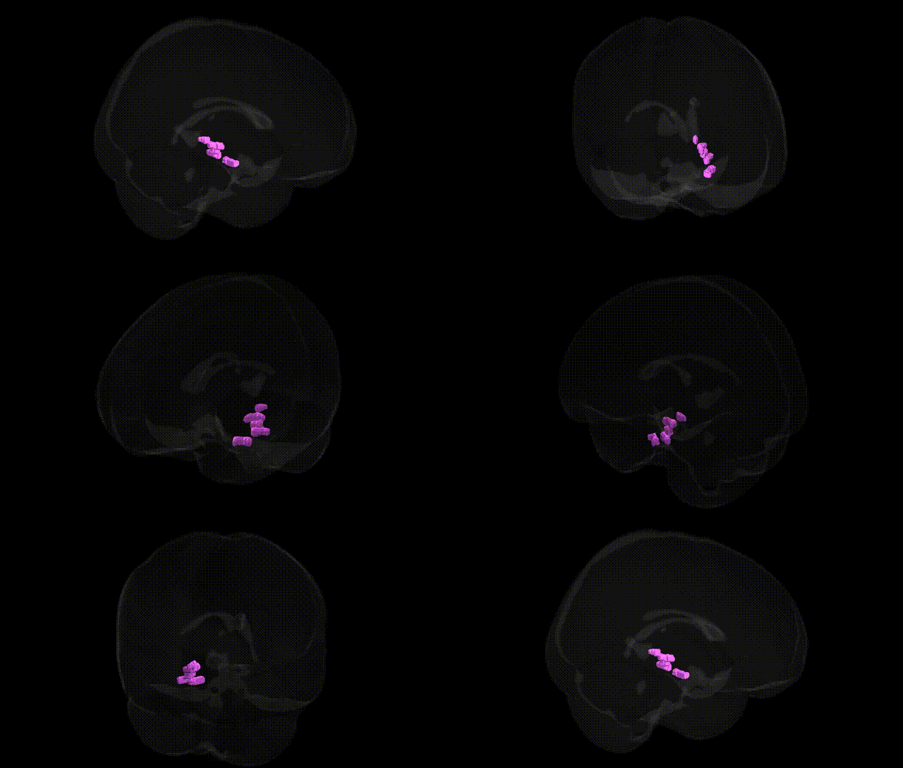
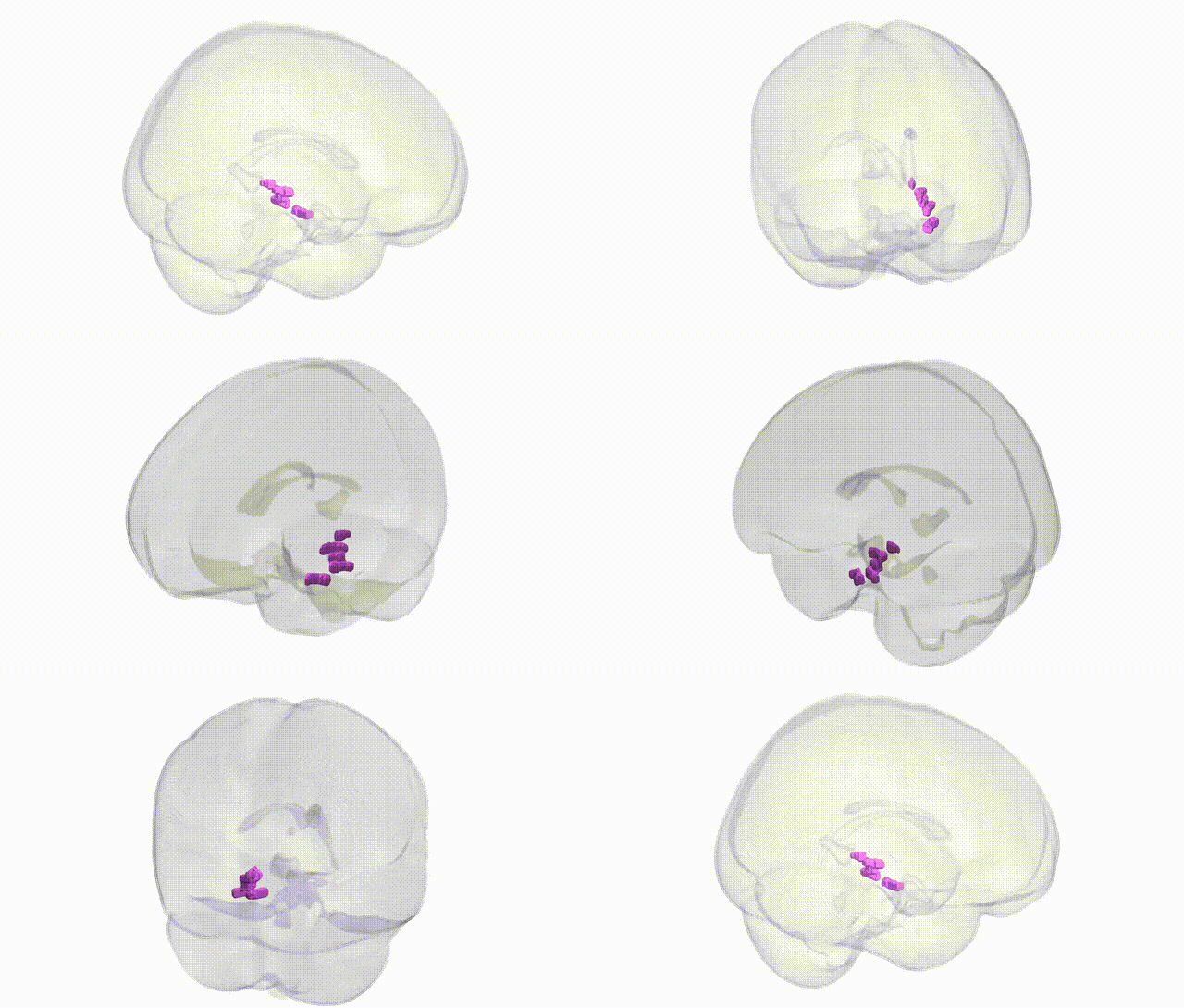
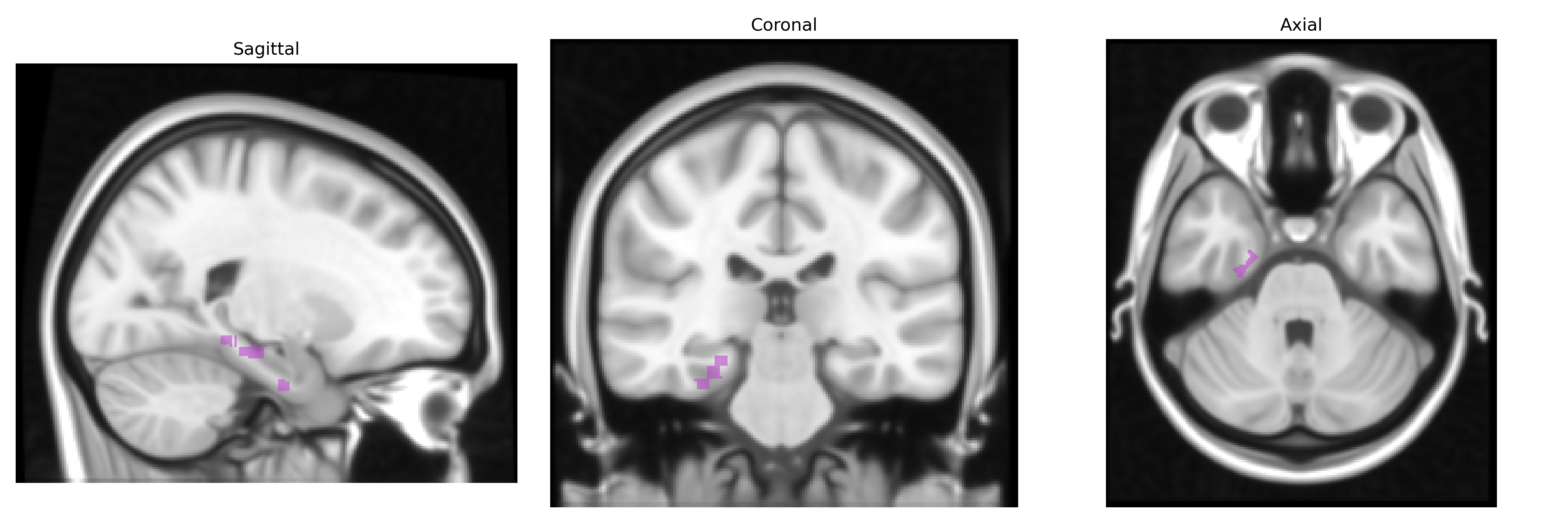
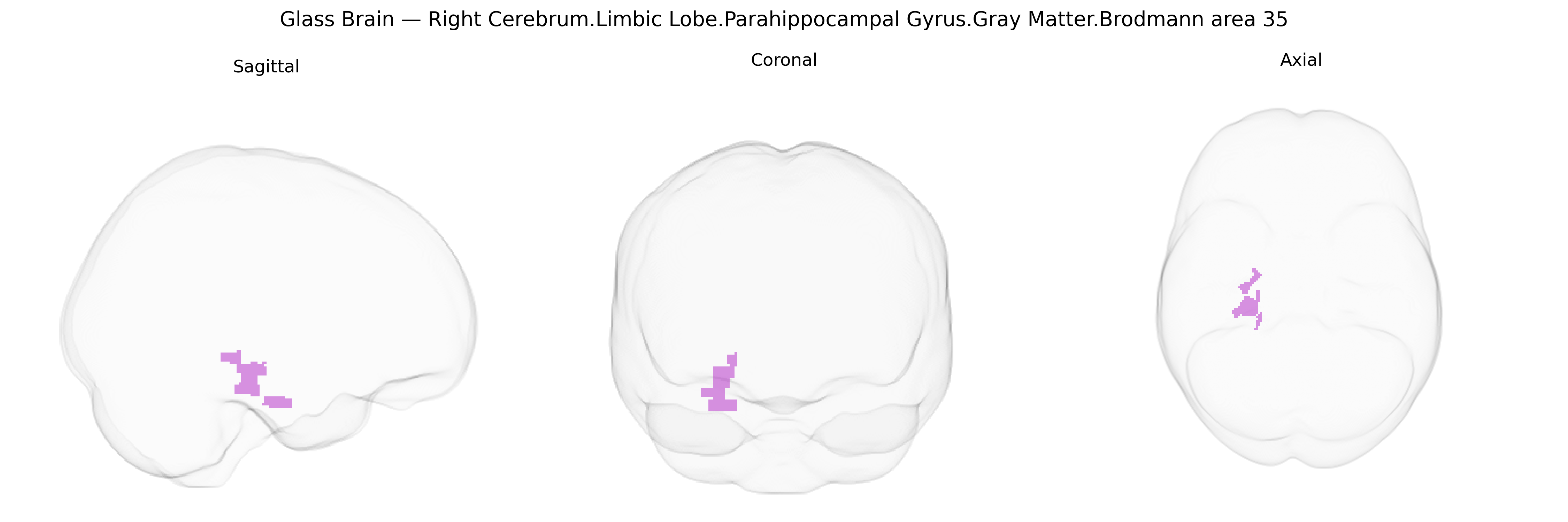
The bilateral Right Cerebrum.Limbic Lobe.Parahippocampal Gyrus.Gray Matter.Brodmann area 35 corresponds to a field in the medial temporal lobe located within the parahippocampal gyrus, forming part of the perirhinal cortex that lies adjacent to the entorhinal cortex and hippocampal formation. Cytoarchitectonically, Brodmann area 35 is characterized by a laminated cortex with features intermediate between classic isocortex and allocortex, reflecting its role as a transitional zone between neocortical association areas and hippocampal structures. Functionally, this area contributes to declarative memory processes, especially familiarity-based recognition and associative memory, and participates in integrating multimodal sensory inputs with mnemonic and emotional context. It forms a key component of the limbic circuitry, with strong connectivity to the hippocampus, amygdala, and higher-order visual areas, and is implicated in early pathological changes in temporal lobe epilepsy and Alzheimer’s disease. There is no direct link for Brodmann area 35, but it is closely related to the Parahippocampal gyrus.
The bilateral right parahippocampal gyrus (PHG) gray matter in Brodmann area 35, as defined in the Talairach 1 mm atlas, has been implicated in numerous imaging genetics and GWAS studies linking genetic variation to limbic and medial temporal lobe structure and function, particularly in relation to memory, emotion, and neurodegeneration. Common variants in APOE (especially ε4) show robust associations with reduced PHG volume, altered connectivity, and increased Alzheimer’s disease risk, while loci in or near CLU, PICALM, CR1, and BIN1 have been associated with medial temporal atrophy and parahippocampal cortical thinning in dementia-related GWAS and endophenotype analyses. Large-scale imaging GWAS (e.g., ENIGMA, UK Biobank) have identified SNPs in genes involved in neurodevelopment, synaptic function, and axonal guidance (such as DLG2, SLC39A8, and multiple 3p, 5q, and 12q loci) associated with PHG cortical thickness and surface area, reflecting polygenic influences on limbic morphology. Schizophrenia and major depression GWAS, including imaging–genetics studies, link risk loci (e.g., CACNA1C, ZNF804A, and several MHC-region variants) to structural and functional changes in parahippocampal and adjacent corticolimbic regions, consistent with PHG involvement in episodic memory, context processing, and emotional regulation. Additional associations include PHG alterations tied to genetic risk scores for autism spectrum disorder, post-traumatic stress disorder, and anxiety-related traits, as well as variants influencing neuroticism and memory performance, underscoring this region’s role as a heritable neuroanatomical substrate bridging genetic risk with cognitive and affective phenotypes.
Overview generated by GPT-4o (2026).
Region ID: 141
Hemisphere: bilateral
Atlas: Talairach labels 1mm
Full Quality Version: Download MP4
Full Quality Version: Download MP4
Wali Sidiqyar*, Gaurav Rudravaram*, Elyssa M. McMaster, Trent M. Schwartz, Adam M. Saunders, Kurt G. Schilling, Bennett A. Landman "Introducing SPINS: A Shared Public Visualization Library of Neuroanatomical Structures." Medical Imaging with Deep Learning- short paper
This resource is licensed under CC0 1.0 Universal (Public Domain).