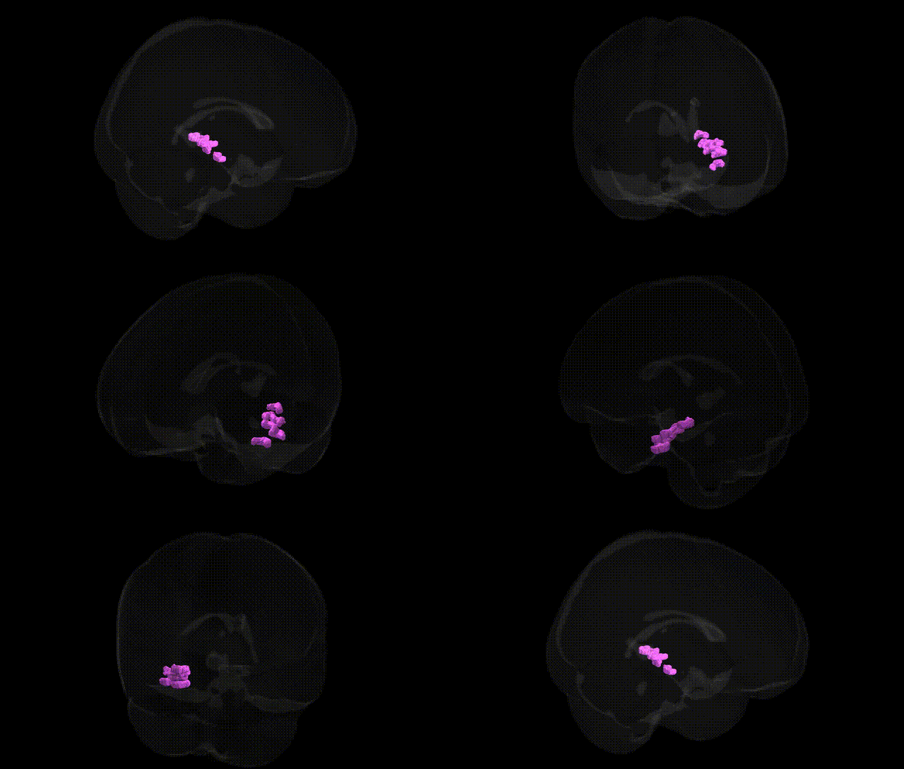
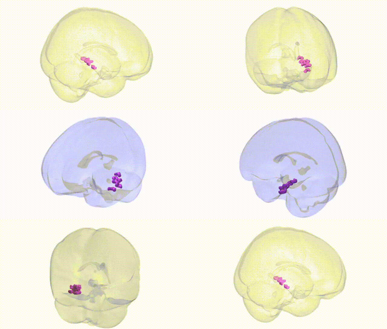
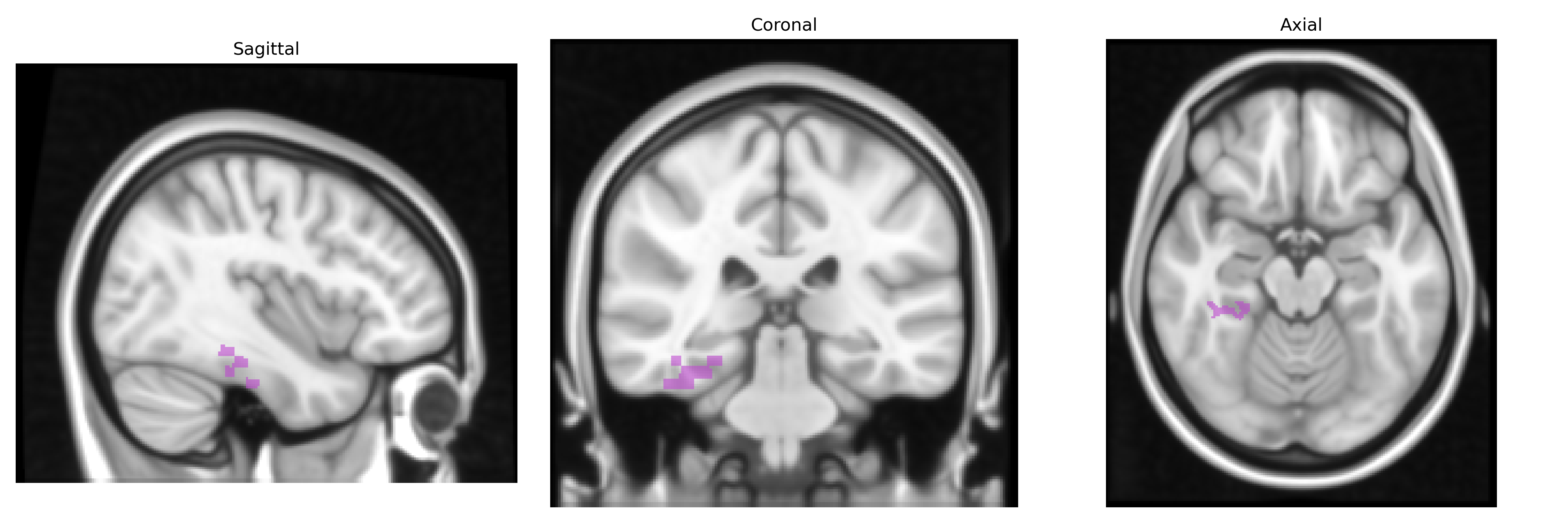
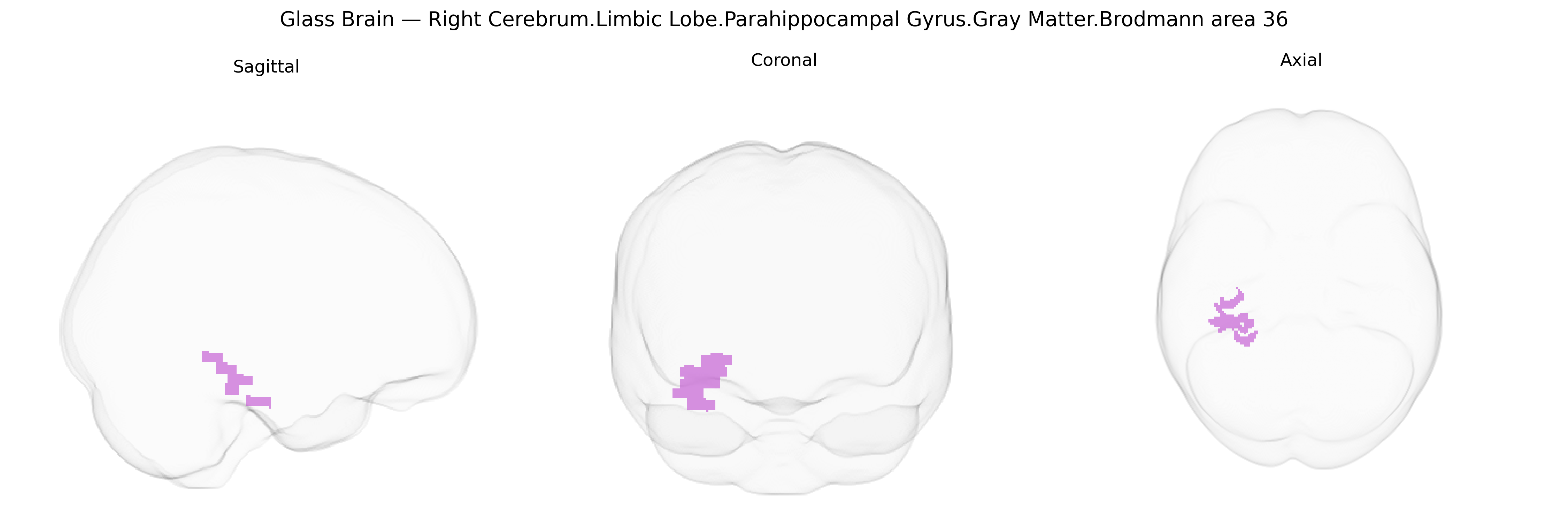
The bilateral Right Cerebrum.Limbic Lobe.Parahippocampal Gyrus.Gray Matter.Brodmann area 36 corresponds to the ectorhinal cortex, a ventromedial temporal lobe region situated along the parahippocampal gyrus, adjacent to the perirhinal (BA35) and entorhinal cortices, and closely associated with the hippocampal formation. This area receives polymodal sensory input and participates in higher-order visual and mnemonic processing, particularly in object recognition, contextual association, and the encoding and retrieval of episodic and semantic memories. Functionally, it serves as an interface between neocortical association areas and the hippocampal circuitry, contributing to the integration of complex sensory information with stored memory representations and playing a role in early stages of neurodegenerative conditions such as Alzheimer’s disease. There is no direct link for Brodmann area 36; a related structure is the Parahippocampal gyrus.
Brodmann area 36 in the parahippocampal gyrus, a key component of the medial temporal lobe memory system, has been implicated in multiple genetic and GWAS findings, although associations are typically reported at the level of medial temporal or parahippocampal structures rather than Talairach BA36 specifically. Variants in APOE (especially ε4), CLU, BIN1, CR1, ABCA7, and other Alzheimer’s disease risk genes have been linked to reduced gray matter volume and altered connectivity in parahippocampal and adjacent fusiform/entorhinal regions, consistent with the early involvement of BA35/36 in neurodegenerative pathology. Large neuroimaging GWAS consortia such as ENIGMA and UK Biobank have identified common variants near genes including KIBRA (WWC1), BDNF, SORL1, and TOMM40 that associate with medial temporal/parahippocampal cortical thickness, surface area, and hippocampal–parahippocampal circuitry measures, often overlapping with genetic architecture of episodic memory performance. Parahippocampal and BA36 activity and structure show heritable variation associated with schizophrenia and major depressive disorder risk loci (e.g., CACNA1C, ZNF804A, GRM3), with imaging-genetic studies reporting links between these variants and abnormal activation during memory or emotional processing tasks. GWAS of anxiety-related traits, PTSD, and neuroticism implicate variants in stress-response and synaptic plasticity genes (FKBP5, NTRK2, and others) that correlate with parahippocampal/BA36 volume or reactivity in fear and context-processing paradigms. Additional associations have been reported between parahippocampal cortical measures and polygenic scores for educational attainment and general cognitive ability, indicating that BA36 structure and function participates in the broader genetic architecture of human memory, learning, and vulnerability to psychiatric and neurodegenerative disorders.
Overview generated by GPT-4o (2026).
Region ID: 130
Hemisphere: bilateral
Atlas: Talairach labels 1mm
Full Quality Version: Download MP4
Full Quality Version: Download MP4
Wali Sidiqyar*, Gaurav Rudravaram*, Elyssa M. McMaster, Trent M. Schwartz, Adam M. Saunders, Kurt G. Schilling, Bennett A. Landman "Introducing SPINS: A Shared Public Visualization Library of Neuroanatomical Structures." Medical Imaging with Deep Learning- short paper
This resource is licensed under CC0 1.0 Universal (Public Domain).