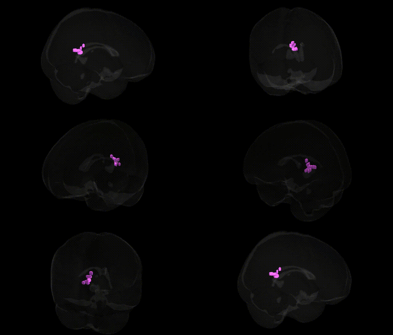
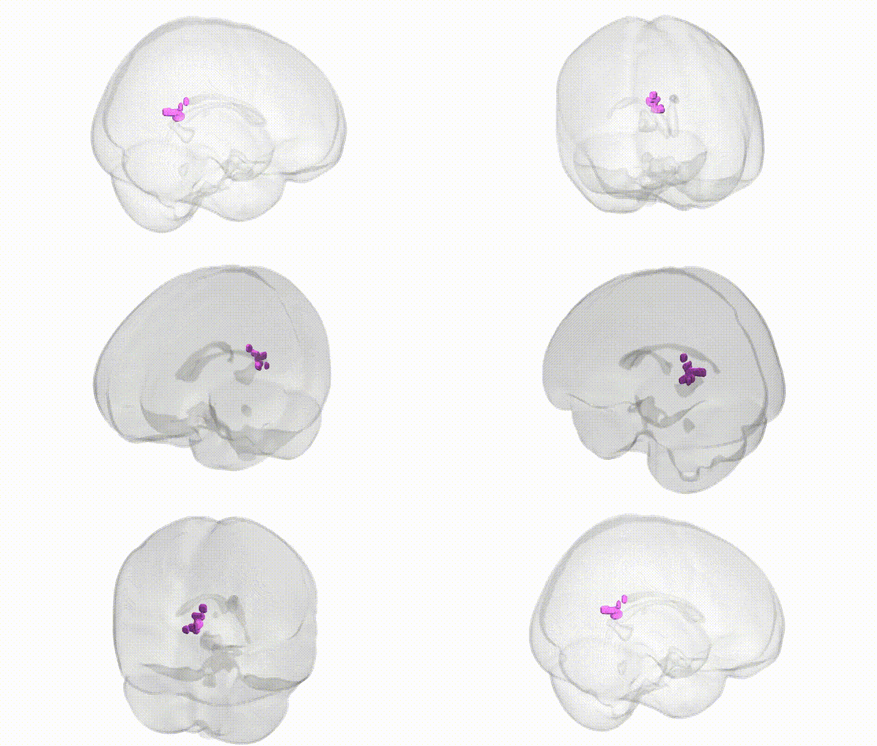
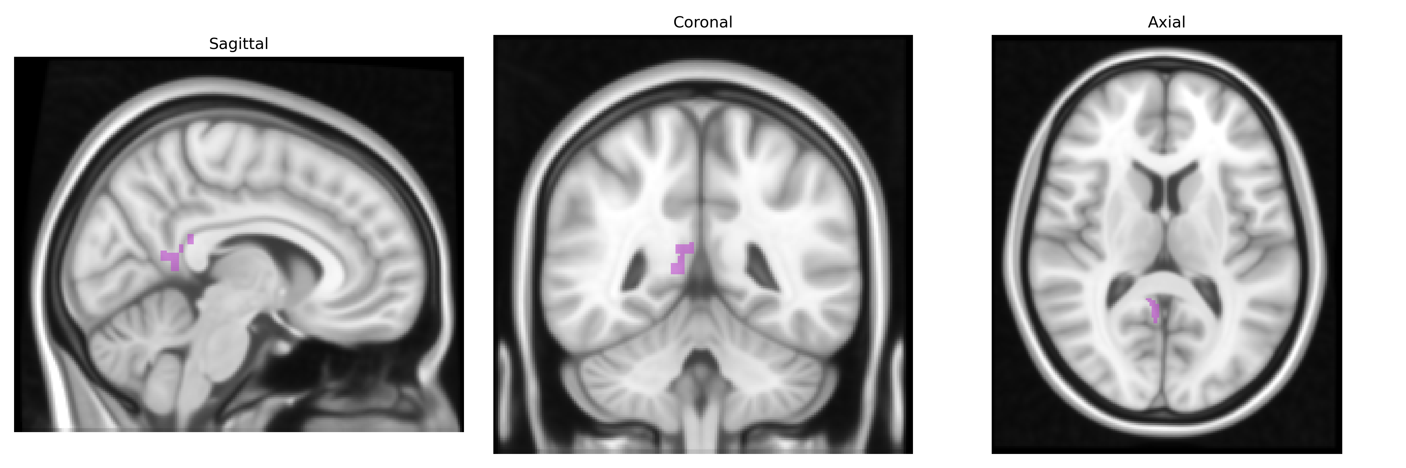
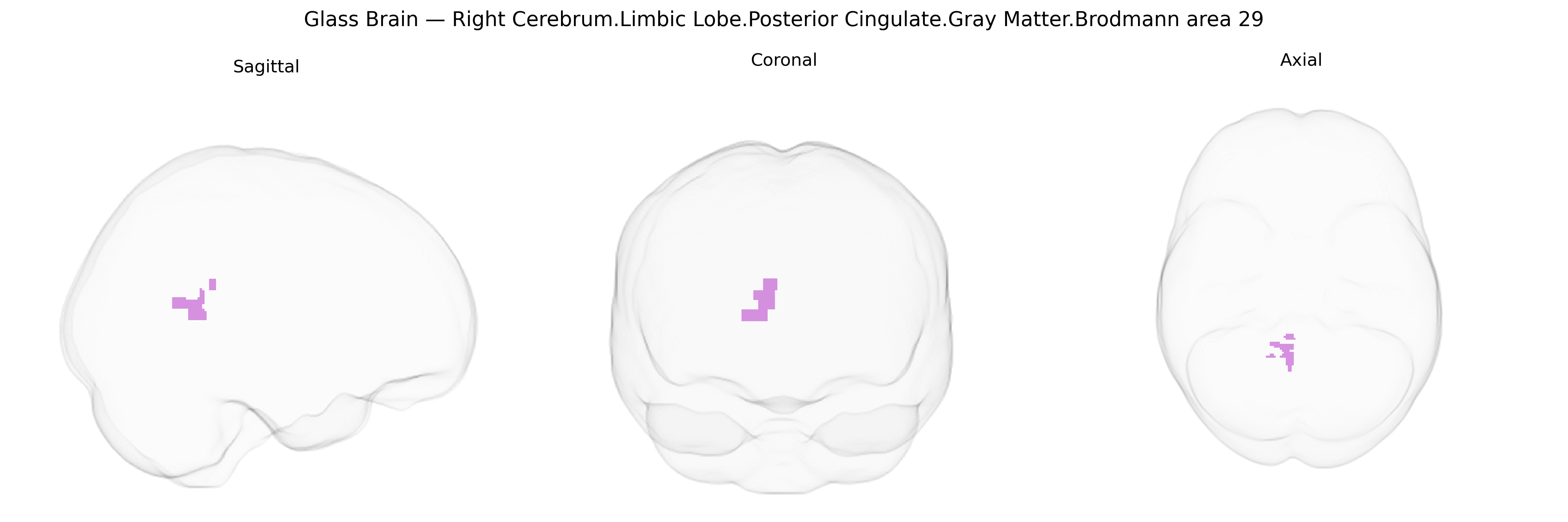
The bilateral Right Cerebrum.Limbic Lobe.Posterior Cingulate.Gray Matter.Brodmann area 29 corresponds to a small agranular cortical field located in the retrosplenial/posterior cingulate region, adjacent to the splenium of the corpus callosum and forming part of the limbic lobe. Brodmann area 29 is characterized by a relatively thin cortical mantle with limbic-type cytoarchitecture and strong connectivity to the hippocampal formation, parahippocampal cortex, and other posterior cingulate and retrosplenial areas, supporting roles in episodic memory, spatial navigation, and contextual processing. Functionally, this region contributes to integrating multimodal sensory and mnemonic information, is often active in tasks involving autobiographical memory and scene construction, and forms part of broader default mode and limbic networks implicated in internal mentation and emotional-cognitive integration. There is no direct link for Brodmann area 29; a related structure is the Posterior cingulate cortex.
Brodmann area 29 (retrosplenial/posterior cingulate cortex within the limbic lobe) shows convergent genetic associations through imaging‑genetics and GWAS linking structural or functional variation in this region to several neuropsychiatric and cognitive traits. Twin and SNP‑heritability studies indicate that posterior cingulate/retrosplenial gray matter volume and connectivity have moderate heritability, with common variants in APOE (especially ε4) and other Alzheimer’s disease risk genes (e.g., CLU, PICALM) repeatedly associated with altered posterior cingulate metabolism, atrophy, and connectivity in preclinical and clinical Alzheimer’s disease. Large neuroimaging GWAS consortia (ENIGMA, UK Biobank) have identified loci in or near genes involved in synaptic function, neurodevelopment, and myelination (such as BDNF, MS4A cluster, and various chromatin‑remodeling genes) that correlate with posterior cingulate thickness or volume, often overlapping with loci implicated in general cognitive ability, educational attainment, and default mode network activity. Genetic risk for major depression, schizophrenia, and autism spectrum disorder, captured by polygenic risk scores, has been associated with abnormal posterior cingulate/retrosplenial structure and functional connectivity, particularly within the default mode network. Additionally, posterior cingulate involvement has been reported in GWAS‑informed studies of PTSD and anxiety, where risk variants in stress‑response and immune‑related genes modulate connectivity between limbic structures and retrosplenial/posterior cingulate cortex. Collectively, these findings position BA29 and adjacent posterior cingulate regions as heritable, genetically modulated hubs linking dementia, psychiatric risk, and individual differences in memory and self‑referential processing.
Overview generated by GPT-4o (2026).
Region ID: 671
Hemisphere: bilateral
Atlas: Talairach labels 1mm
Full Quality Version: Download MP4
Full Quality Version: Download MP4
Wali Sidiqyar*, Gaurav Rudravaram*, Elyssa M. McMaster, Trent M. Schwartz, Adam M. Saunders, Kurt G. Schilling, Bennett A. Landman "Introducing SPINS: A Shared Public Visualization Library of Neuroanatomical Structures." Medical Imaging with Deep Learning- short paper
This resource is licensed under CC0 1.0 Universal (Public Domain).