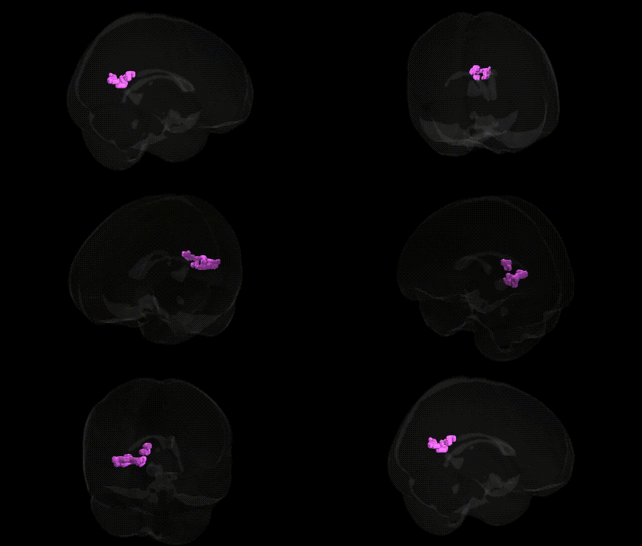
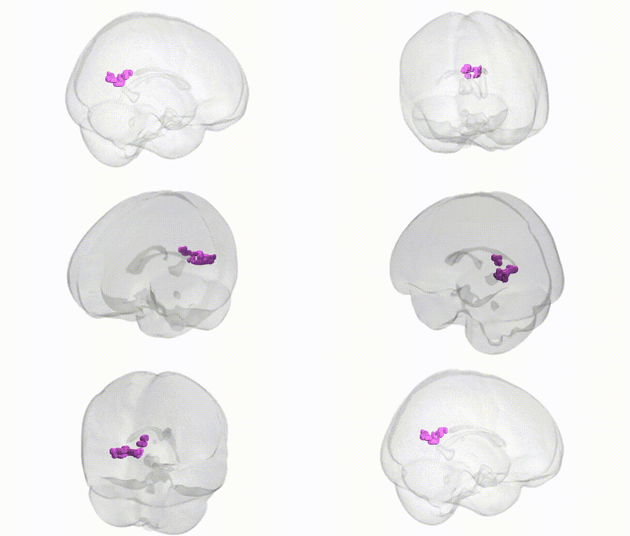
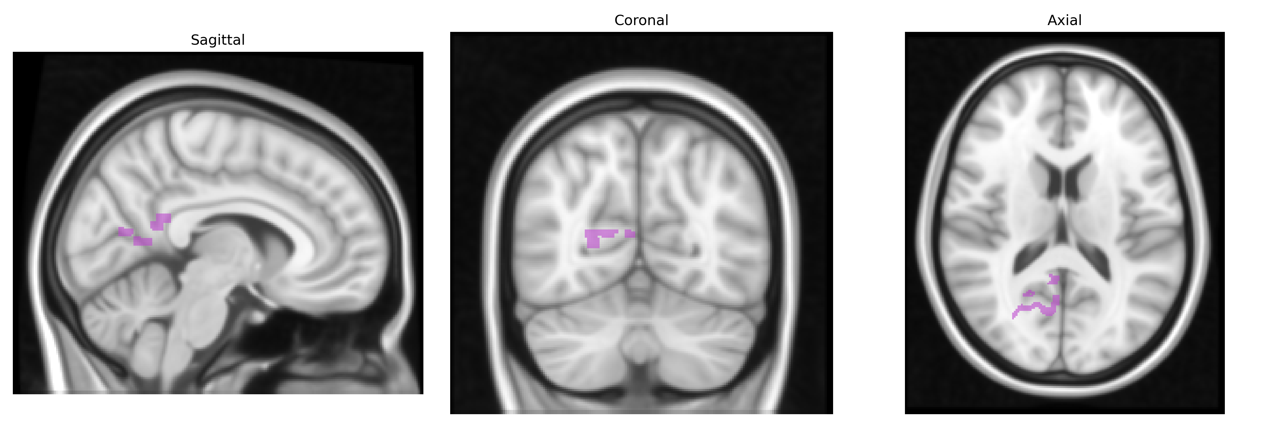
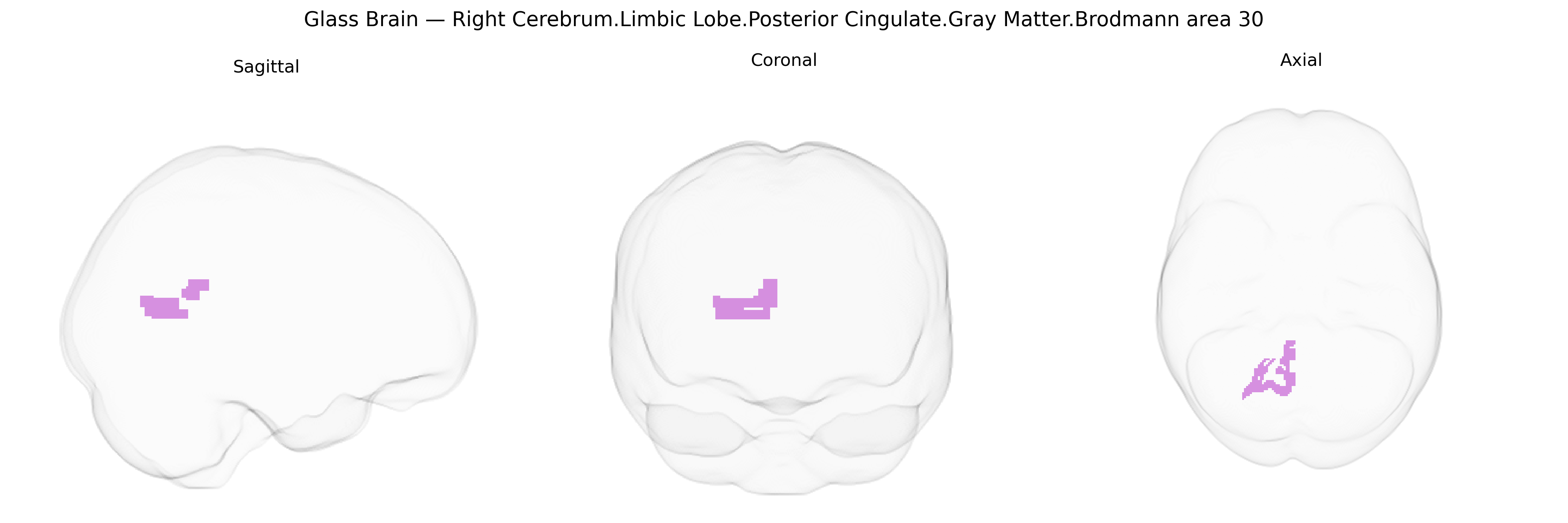
The bilateral Right Cerebrum.Limbic Lobe.Posterior Cingulate.Gray Matter.Brodmann area 30 corresponds to a caudal subdivision of the posterior cingulate cortex, located on the medial aspect of the parietal lobe within the limbic network. Brodmann area 30 (the agranular retrolimbic area) lies ventral and posterior to BA23 and BA31, bordering the retrosplenial cortex and closely associated with the cingulate gyrus and corpus callosum. Cytoarchitectonically, it is characterized by a less distinct granular layer and limbic-type cortical organization, reflecting strong connections with the hippocampal formation, parahippocampal regions, thalamus, and other default mode network nodes. Functionally, this region is implicated in internally directed cognition, autobiographical and episodic memory, spatial and contextual processing, and integration of emotional and mnemonic information, contributing to self-referential processing and baseline brain activity. There is no direct link for Brodmann area 30; a closely related structure is the Posterior cingulate cortex.
The bilateral right posterior cingulate cortex (PCC) in Brodmann area 30, part of the limbic lobe gray matter, has been repeatedly implicated in genetic and genome-wide association studies (GWAS) of brain structure, functional connectivity, and neuropsychiatric disease, although many findings involve broader PCC or posterior default mode network regions rather than BA30 alone. Imaging-genetics work shows heritable variation in PCC thickness, surface area, and resting-state connectivity, with polygenic influences overlapping those for general cognitive ability, educational attainment, and Alzheimer’s disease (AD). Large GWAS of cortical morphology (e.g., ENIGMA and UK Biobank) have identified common variants in genes related to neurodevelopment, synaptic function, and myelination (such as variants near genes like MIR2113, HMGA2, and others) associated with posterior cingulate or adjacent precuneus morphology. AD GWAS and endophenotype studies highlight the PCC as a key node where APOE ε4 and other AD risk variants (e.g., in CLU, PICALM, CR1) modulate amyloid burden, hypometabolism, and atrophy; PCC alterations also appear in polygenic risk score analyses for AD and dementia. Genetic influences on PCC structure and activity have additionally been linked to major depressive disorder, schizophrenia, autism spectrum disorder, and attention-deficit/hyperactivity disorder via large-scale imaging-genetics consortia, often through shared polygenic architecture rather than single-locus effects. While specific BA30-focused GWAS are limited, converging evidence indicates that posterior cingulate regions, including BA30, serve as structurally and functionally heritable hubs where common genetic variation contributes to risk for neurodegenerative disease, psychiatric disorders, and individual differences in cognition and default mode network function.
Overview generated by GPT-4o (2026).
Region ID: 665
Hemisphere: bilateral
Atlas: Talairach labels 1mm
Full Quality Version: Download MP4
Full Quality Version: Download MP4
Wali Sidiqyar*, Gaurav Rudravaram*, Elyssa M. McMaster, Trent M. Schwartz, Adam M. Saunders, Kurt G. Schilling, Bennett A. Landman "Introducing SPINS: A Shared Public Visualization Library of Neuroanatomical Structures." Medical Imaging with Deep Learning- short paper
This resource is licensed under CC0 1.0 Universal (Public Domain).