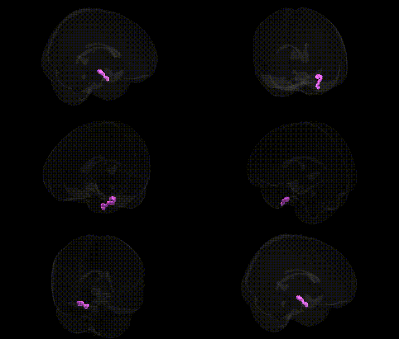
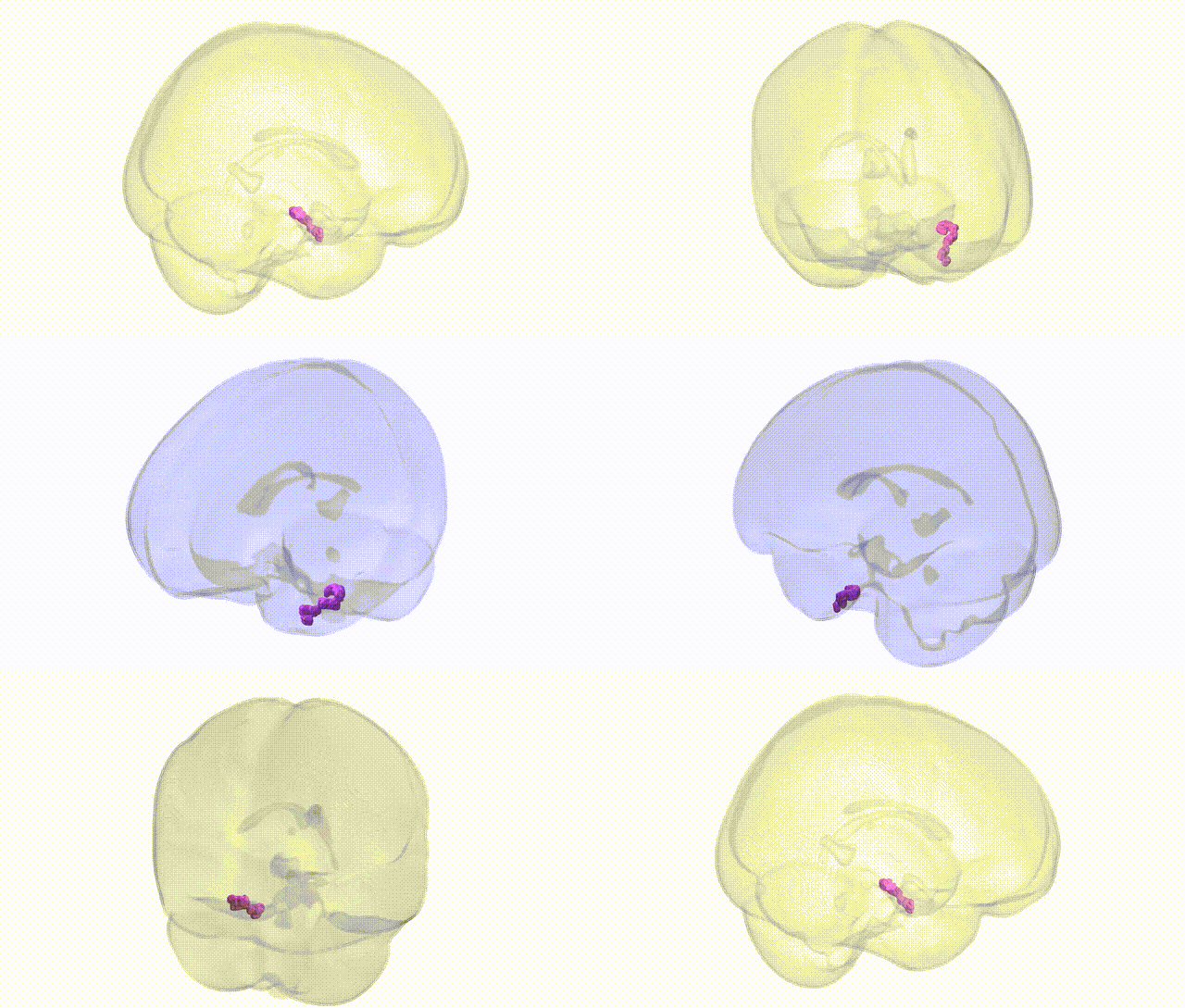
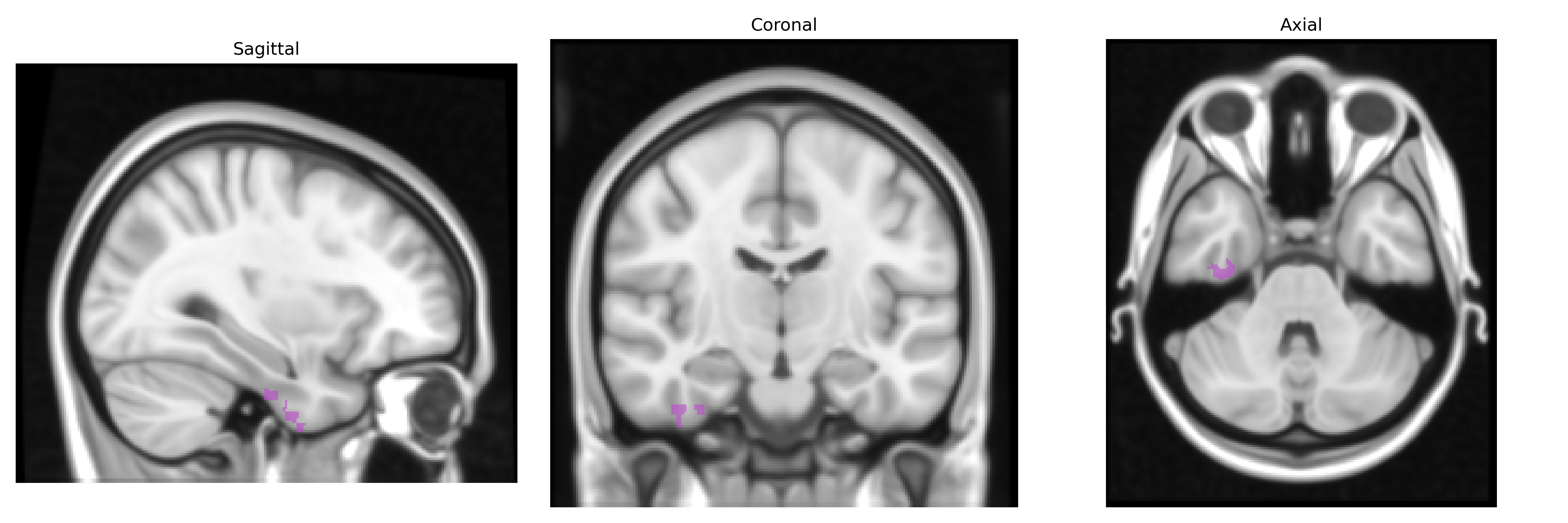
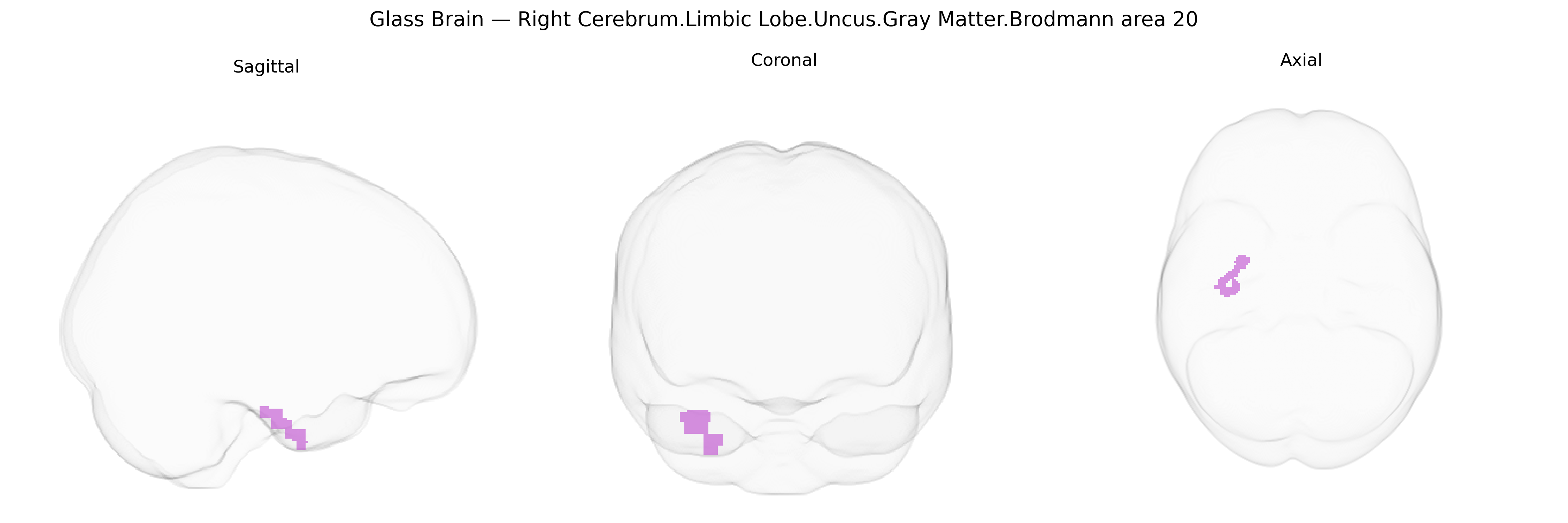
The bilateral Right Cerebrum.Limbic Lobe.Uncus.Gray Matter.Brodmann area 20 corresponds to portions of the anterior medial temporal cortex where the uncus, a medial protrusion of the parahippocampal gyrus overlying the amygdaloid complex, overlaps with the ventral inferotemporal region assigned to Brodmann area 20. This cortex is composed of association-type gray matter involved in higher-order visual object processing, semantic integration, and limbic-related functions such as emotional evaluation and memory-related context encoding. Its position along the medial temporal surface links visual input from inferotemporal pathways with limbic structures (hippocampus and amygdala), supporting the integration of perceptual information with affective and mnemonic processes. There is no direct Wikipedia article for this exact composite label; a closely related structure is the Uncus.
The bilateral right cerebrum limbic lobe uncus in Brodmann area 20 lies at the interface of medial temporal and ventral visual association cortex, so genetic associations typically emerge from studies of medial temporal, anterior parahippocampal, and ventral temporal regions rather than this exact Talairach-defined parcel. GWAS and imaging-genetics studies implicate variants in genes involved in synaptic function and neurodevelopment—such as BDNF (e.g., Val66Met), APOE (particularly ε4), and several schizophrenia and autism risk loci—in structural and functional changes spanning the medial temporal lobe, including adjacent uncus and parahippocampal regions, where BA20 contributes to higher-order visual and semantic processing. APOE ε4 carrier status is consistently associated with gray matter loss and altered functional connectivity in medial temporal and limbic areas relevant to BA20 and uncus, in the context of Alzheimer’s disease and age-related cognitive decline. Polygenic risk scores for major depressive disorder, bipolar disorder, schizophrenia, and autism spectrum disorder have been linked to volume and connectivity changes in overlapping limbic and ventral temporal networks, while GWAS of brain imaging phenotypes (e.g., ENIGMA, UK Biobank) identify multiple common variants—often in genes regulating neuronal development, axon guidance, and synaptic plasticity—that influence cortical thickness and surface area in temporal association cortices encompassing BA20. However, no large-scale study has isolated genetic associations specific to the right BA20 uncus gray matter parcel, so existing findings reflect broader medial temporal and ventral temporal genetic influences rather than a region-exclusive profile.
Overview generated by GPT-4o (2026).
Region ID: 18
Hemisphere: bilateral
Atlas: Talairach labels 1mm
Full Quality Version: Download MP4
Full Quality Version: Download MP4
Wali Sidiqyar*, Gaurav Rudravaram*, Elyssa M. McMaster, Trent M. Schwartz, Adam M. Saunders, Kurt G. Schilling, Bennett A. Landman "Introducing SPINS: A Shared Public Visualization Library of Neuroanatomical Structures." Medical Imaging with Deep Learning- short paper
This resource is licensed under CC0 1.0 Universal (Public Domain).