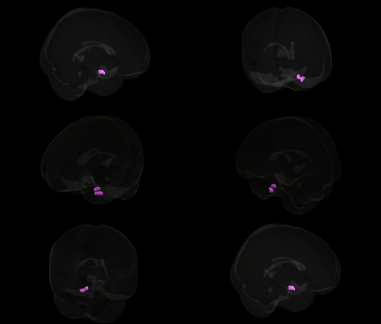
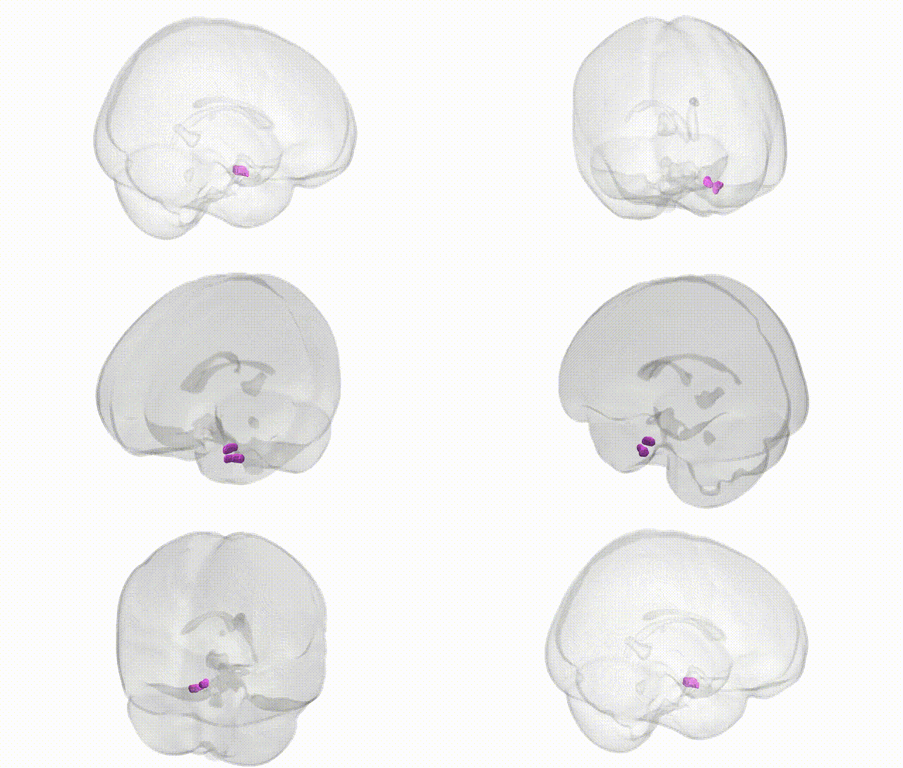
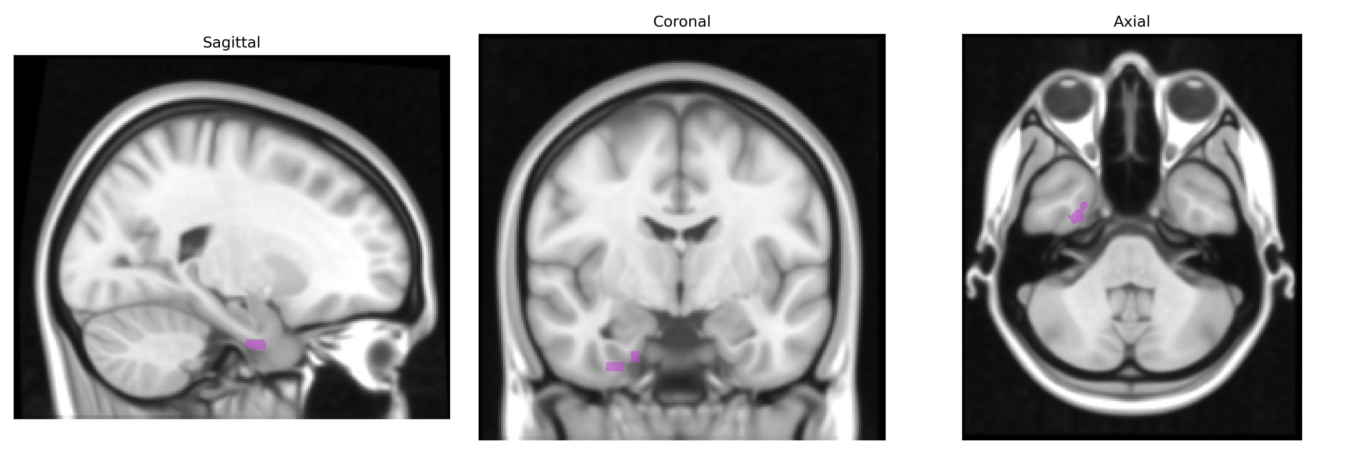
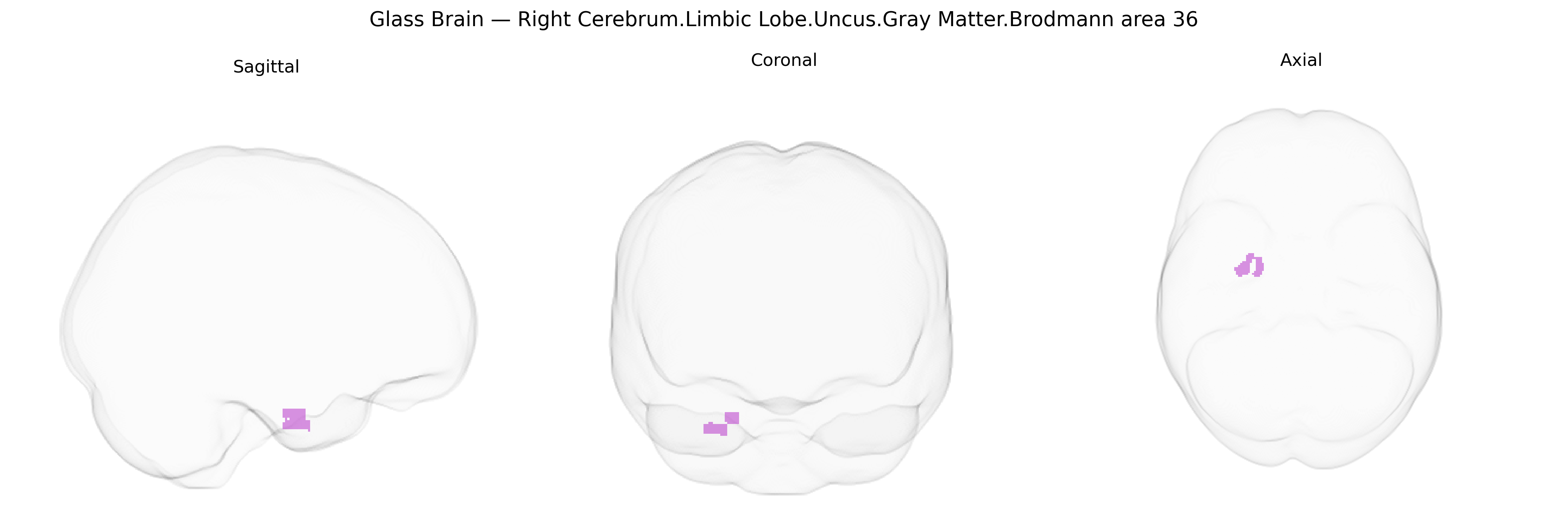
The bilateral Right Cerebrum.Limbic Lobe.Uncus.Gray Matter.Brodmann area 36 corresponds to the perirhinal cortex region of the medial temporal lobe, situated in the anterior parahippocampal gyrus and closely associated with the uncus and amygdalo-hippocampal complex. Architectonically, Brodmann area 36 is agranular to dysgranular cortex, forming part of the parahippocampal–entorhinal–hippocampal circuitry critical for object recognition memory, associative learning, and higher-order integration of multimodal sensory information. It receives convergent visual and polymodal inputs from temporal association cortices and projects to entorhinal cortex and hippocampus, contributing to familiarity-based recognition and the encoding and retrieval of episodic-like memory traces. Pathologically, this region is frequently involved in early neurodegenerative changes in medial temporal lobe epilepsy and in prodromal Alzheimer’s disease, reflecting its central role in memory-related limbic networks. There is no direct link for Brodmann area 36; a closely related structure is the Perirhinal cortex.
The bilateral right Brodmann area 36 in the uncus of the limbic lobe, typically mapped within parahippocampal and perirhinal cortex territories in the Talairach 1 mm atlas, is implicated by genetic and GWAS-based imaging genetics studies primarily through associations with episodic memory, recognition memory, and medial temporal lobe–dependent learning, as well as with risk for neuropsychiatric and neurodegenerative disorders. Polygenic risk scores for Alzheimer’s disease, schizophrenia, and major depressive disorder have been associated with structural and functional alterations in adjacent medial temporal and parahippocampal regions encompassing BA36, with notable contributions from loci in APOE, CLU, PICALM, BIN1, and other Alzheimer’s-related genes, and from schizophrenia risk loci such as those in the MHC region and CACNA1C that modulate medial temporal volume and connectivity. Variants in genes involved in synaptic plasticity (e.g., BDNF Val66Met), glutamatergic and GABAergic signaling, and neurodevelopmental pathways (including some 22q11.2 deletion–related genes) have been linked to differences in medial temporal and parahippocampal gray matter volumes and activation during memory tasks that recruit BA36. Large-scale brain imaging GWAS consortia (e.g., ENIGMA, UK Biobank) report that common variants exert small but significant effects on parahippocampal and entorhinal thickness and volume—measures that often include or border BA36—supporting a highly polygenic architecture in which many loci of modest effect influence the structure and function of this region and contribute to susceptibility for dementia, mood and psychotic disorders, and individual variability in memory-related traits.
Overview generated by GPT-4o (2026).
Region ID: 76
Hemisphere: bilateral
Atlas: Talairach labels 1mm
Full Quality Version: Download MP4
Full Quality Version: Download MP4
Wali Sidiqyar*, Gaurav Rudravaram*, Elyssa M. McMaster, Trent M. Schwartz, Adam M. Saunders, Kurt G. Schilling, Bennett A. Landman "Introducing SPINS: A Shared Public Visualization Library of Neuroanatomical Structures." Medical Imaging with Deep Learning- short paper
This resource is licensed under CC0 1.0 Universal (Public Domain).