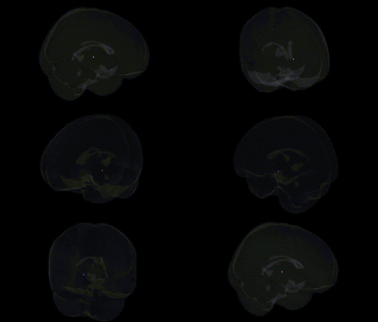
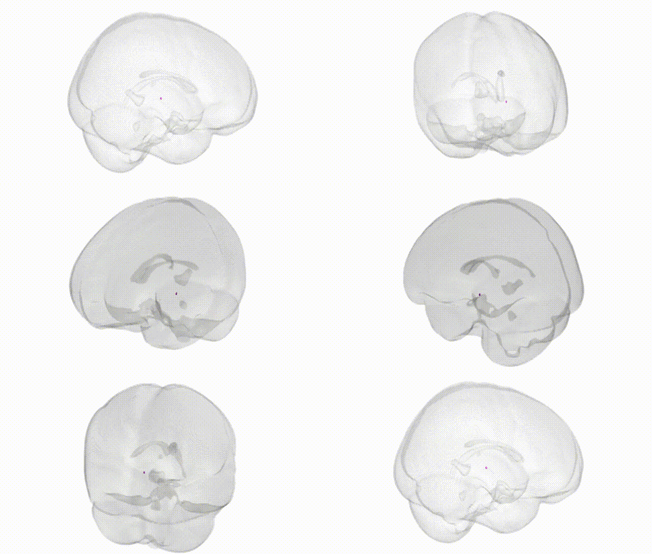
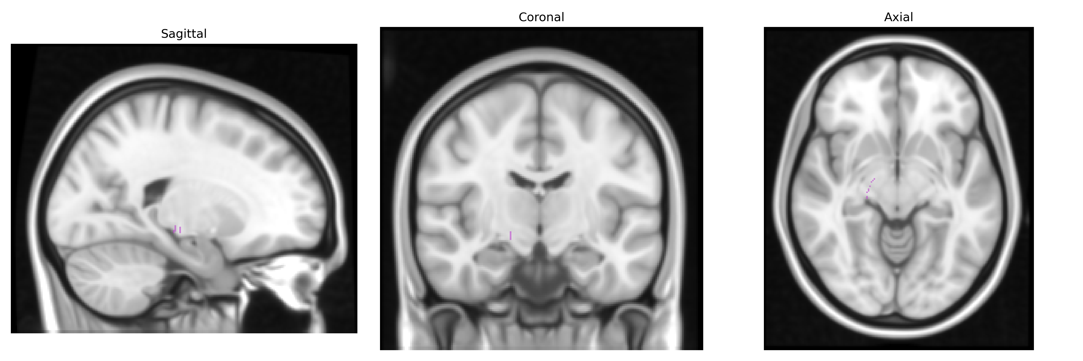
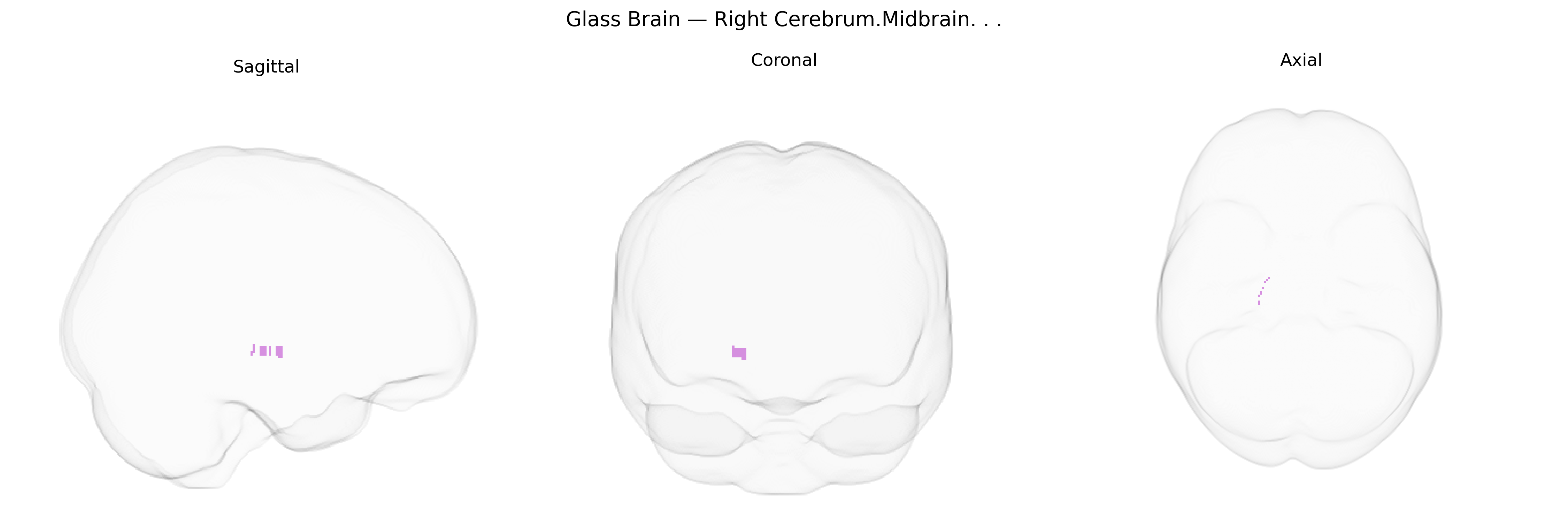
The bilateral Right Cerebrum.Midbrain in the Talairach 1 mm Atlas corresponds to midbrain structures located within the right cerebral hemisphere reference frame, encompassing portions of the mesencephalon involved in sensorimotor integration, eye movement control, arousal, and auditory and visual reflex processing. This region contains nuclei and conduits such as the superior and inferior colliculi (for visual and auditory orienting reflexes), the substantia nigra (critical for dopaminergic modulation of movement and implicated in Parkinson’s disease), and the red nucleus (involved in motor coordination), along with ascending sensory pathways and descending motor tracts that pass between the forebrain and brainstem. Functionally, it serves as a key relay and integration hub linking cortical, cerebellar, and lower brainstem structures, supporting coordinated movement, reflexive responses to sensory stimuli, and regulation of consciousness and sleep–wake states. There is no direct Wikipedia article for “Right Cerebrum.Midbrain” as a Talairach label; a related structure is the Midbrain.
The bilateral right midbrain, as defined in the Talairach 1 mm atlas, encompasses dopaminergic nuclei (notably the substantia nigra and ventral tegmental area) and other midbrain structures that have been repeatedly implicated in genetic studies of movement, psychiatric, and neurodevelopmental disorders. GWAS and candidate gene studies link variation in genes affecting dopaminergic signaling and midbrain development—such as SNCA, LRRK2, GBA, PARK2, and MAPT—to Parkinson’s disease, which shows marked neurodegeneration of midbrain dopaminergic neurons, particularly in the substantia nigra. Schizophrenia and other psychotic disorders show genetic overlap in pathways involving dopamine, glutamate, and neuronal development (e.g., DRD2, CACNA1C, GRIN2A, and broader polygenic risk scores), with imaging–genetic work associating these variants with altered midbrain activation and connectivity in cortico-striato-thalamo-midbrain circuits. Midbrain volume and integrity have also been associated with genetic risk for major depressive disorder, bipolar disorder, and addiction-related traits, often via polygenic influences on reward and salience networks, as well as with genes regulating neurodevelopment and axon guidance (e.g., NRG1, SEMA family genes). Neuroimaging–GWAS consortia (such as ENIGMA and UK Biobank) report that common variants distributed across the genome contribute to individual differences in midbrain volume, microstructure, and functional responses, implicating pathways related to synaptic plasticity, mitochondrial function, and neuroinflammation; however, associations are typically polygenic and shared with broader subcortical and brainstem regions rather than being specific to the right midbrain alone.
Overview generated by GPT-4o (2026).
Region ID: 440
Hemisphere: bilateral
Atlas: Talairach labels 1mm
Full Quality Version: Download MP4
Full Quality Version: Download MP4
Wali Sidiqyar*, Gaurav Rudravaram*, Elyssa M. McMaster, Trent M. Schwartz, Adam M. Saunders, Kurt G. Schilling, Bennett A. Landman "Introducing SPINS: A Shared Public Visualization Library of Neuroanatomical Structures." Medical Imaging with Deep Learning- short paper
This resource is licensed under CC0 1.0 Universal (Public Domain).