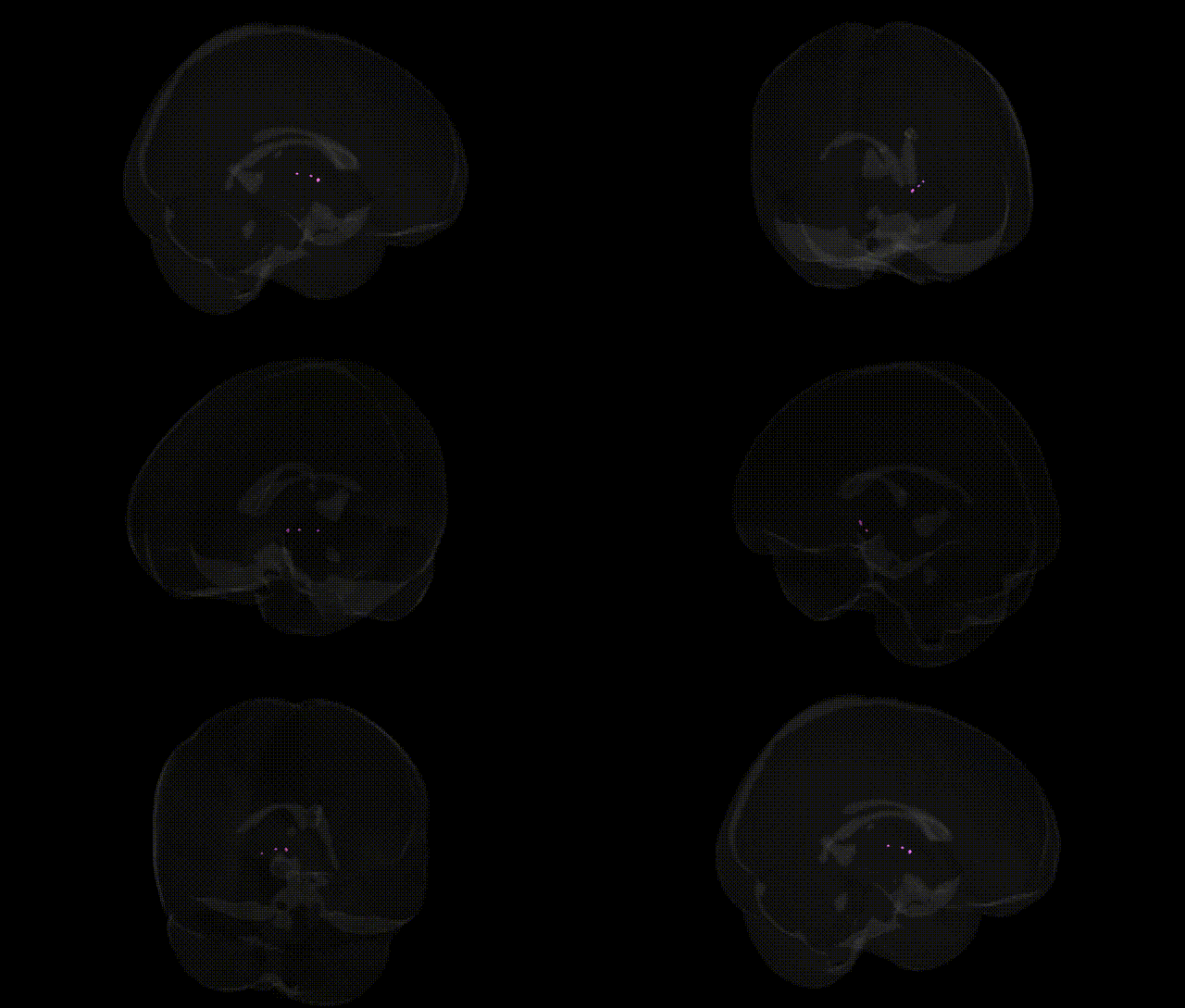
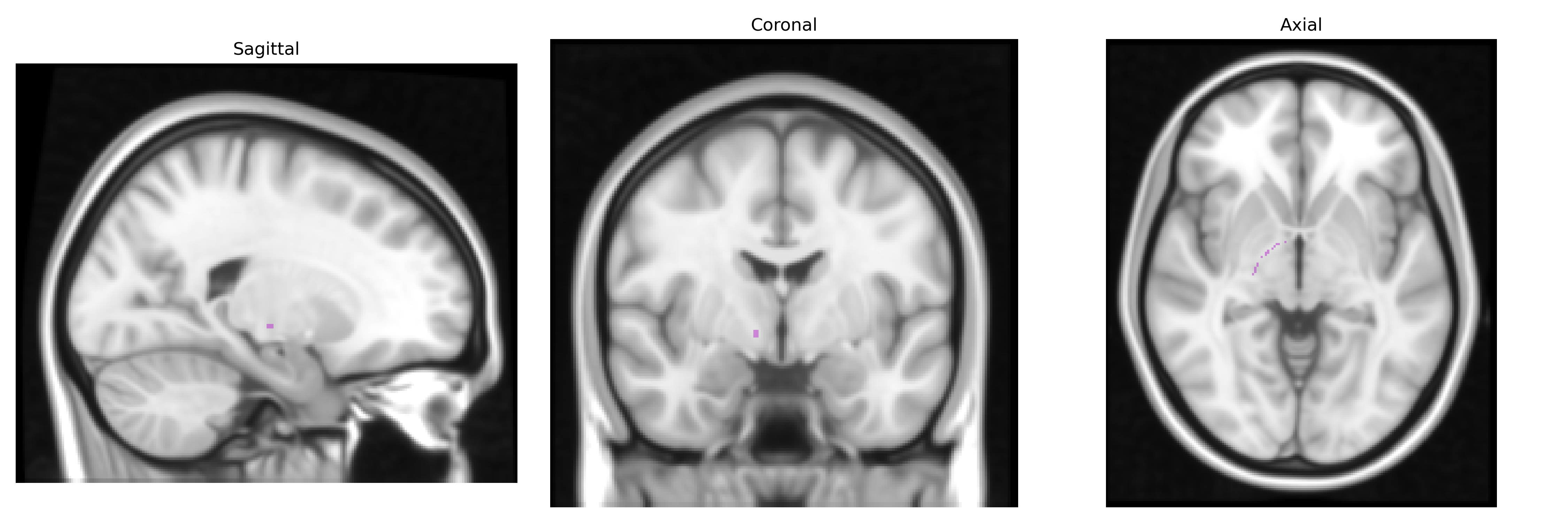
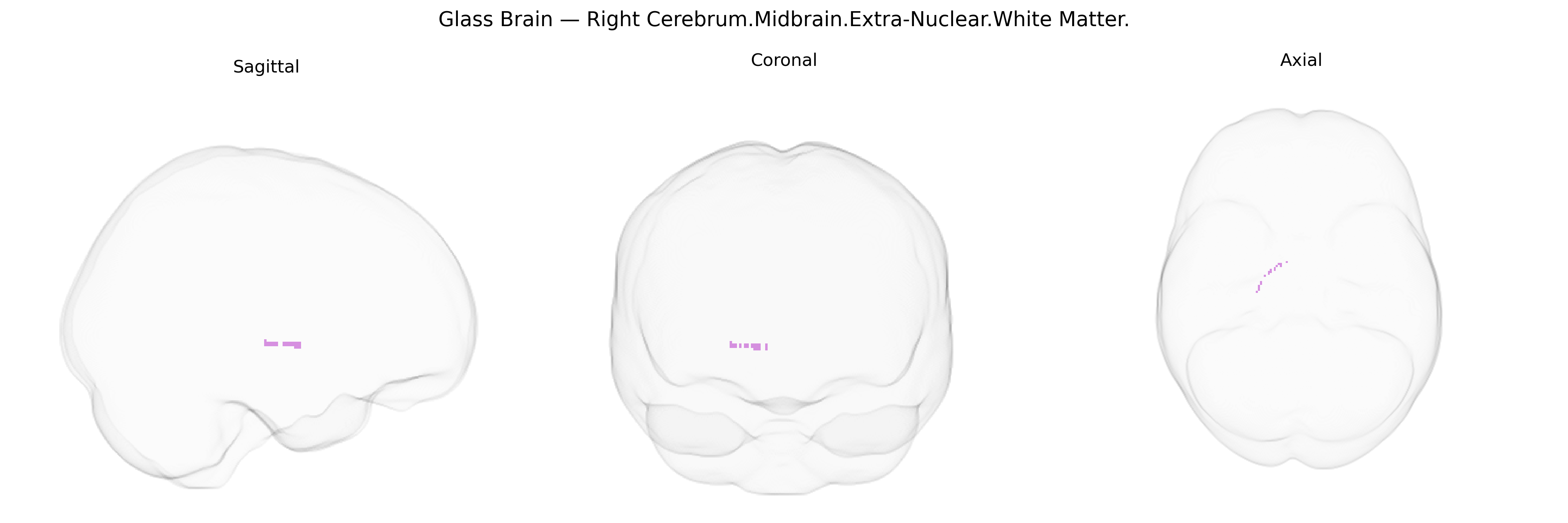
The bilateral Right Cerebrum.Midbrain.Extra-Nuclear.White Matter region in the Talairach 1 mm atlas refers to deep, non-nuclear white matter tracts located within the midbrain portion of the right cerebral hemisphere, excluding the distinct gray matter nuclei such as the substantia nigra and red nucleus. This “extra-nuclear” designation highlights bundles of myelinated axons that conduct signals between cortical areas, brainstem nuclei, and spinal pathways, including components of ascending sensory and descending motor systems coursing through the midbrain tegmentum and surrounding structures. Functionally, these fibers participate in integration of motor control, sensory processing, arousal, and reflexive orienting by linking the cerebral cortex with midbrain centers and lower brainstem-spinal circuits. There is no direct link for this exact atlas label; a closely related structure is the Midbrain.
The bilateral Right Cerebrum.Midbrain.Extra-Nuclear.White Matter label in the Talairach 1 mm atlas corresponds broadly to midbrain white matter tracts rather than a functionally discrete nucleus, so genetic associations derive from studies of midbrain structure, white matter integrity, and related circuits rather than from this exact atlas label. GWAS of brain imaging phenotypes (e.g., UK Biobank) have identified multiple loci influencing midbrain and brainstem volumes and white matter microstructure—most prominently variants near genes involved in axon guidance and myelination such as CNTN4, ROBO1/2, LINGO1, and genes in oligodendrocyte and myelin pathways (e.g., MAG, MOBP). Midbrain white matter is integral to dopaminergic and motor pathways, and genetic studies of Parkinson’s disease (e.g., SNCA, LRRK2, GBA variants) and schizophrenia and other psychiatric disorders (e.g., DRD2, CACNA1C, GRIN2A) implicate networks that include midbrain tracts, with imaging–genetics work showing that risk alleles can modulate diffusion MRI measures and connectivity in these regions. Neurodevelopmental and neurodegenerative GWAS (for traits such as cognitive performance, ADHD, autism, and general brain volume) have also identified polygenic influences on brainstem and midbrain structures, although associations are typically reported at the level of broader brainstem or whole-brain white matter metrics rather than specifically the “Extra-Nuclear” midbrain white matter defined by Talairach labeling.
Overview generated by GPT-4o (2026).
Region ID: 502
Hemisphere: bilateral
Atlas: Talairach labels 1mm
Full Quality Version: Download MP4
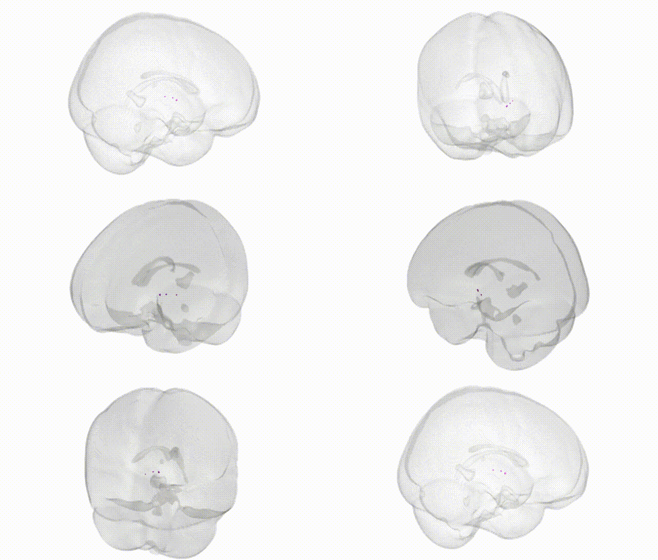
Full Quality Version: Download MP4
Wali Sidiqyar*, Gaurav Rudravaram*, Elyssa M. McMaster, Trent M. Schwartz, Adam M. Saunders, Kurt G. Schilling, Bennett A. Landman "Introducing SPINS: A Shared Public Visualization Library of Neuroanatomical Structures." Medical Imaging with Deep Learning- short paper
This resource is licensed under CC0 1.0 Universal (Public Domain).