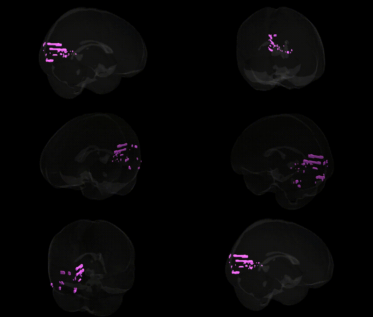
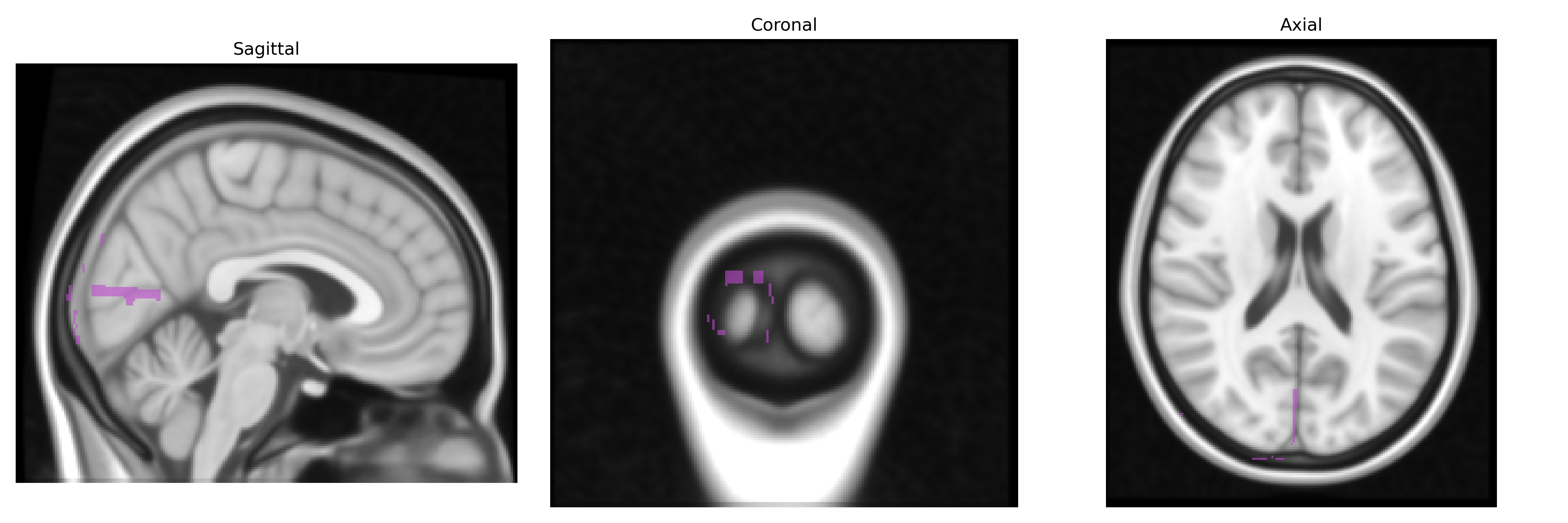
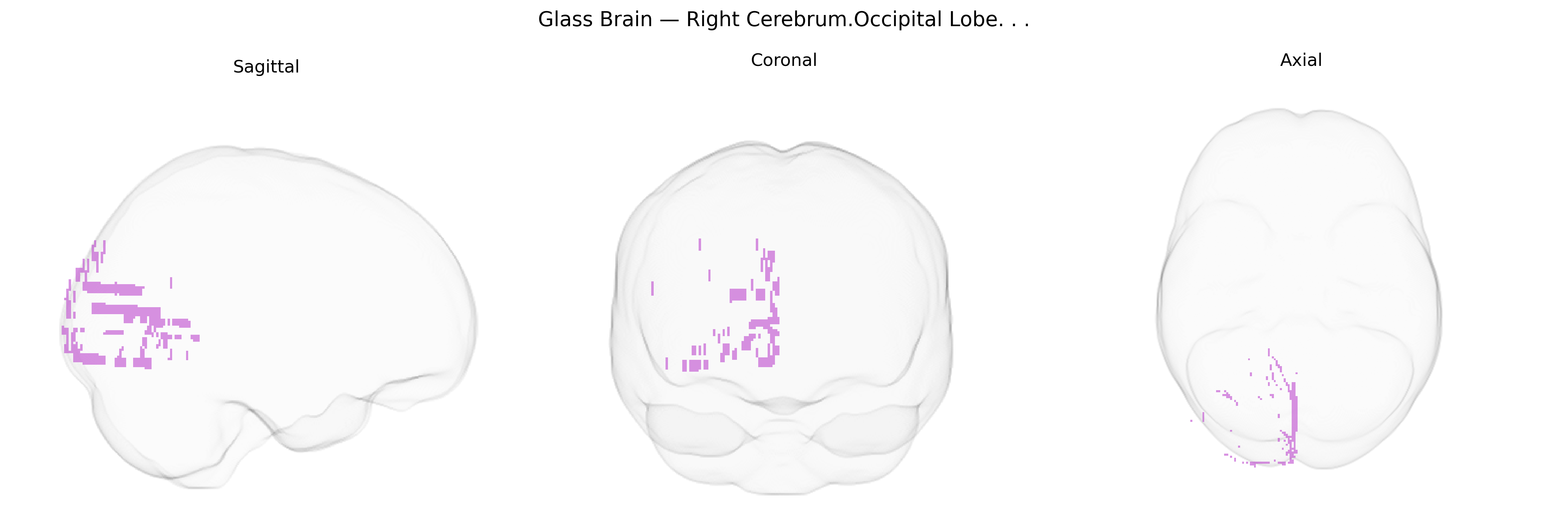
The bilateral Right Cerebrum, Occipital Lobe comprises the posterior cortical territories of the right cerebral hemisphere responsible primarily for visual processing, including the reception, integration, and interpretation of visual stimuli conveyed from the retina via the lateral geniculate nucleus of the thalamus. This lobe contains primary and secondary visual areas (notably Brodmann area 17 and associated extrastriate regions) that support fundamental functions such as object recognition, spatial orientation, motion perception, and color discrimination, while also contributing to visuospatial attention and the integration of visual information with higher-order cognitive processes. Lesions in this region can result in contralateral visual field deficits (e.g., left homonymous hemianopia), visual agnosias, or impairments in complex visuoperceptual tasks, reflecting its essential role in conscious visual experience and visually guided behavior. Occipital lobe
The bilateral right occipital lobe, encompassing primary and associative visual cortex, has been implicated in multiple genetic association studies, particularly GWAS of brain structure and neuropsychiatric traits. Variants near genes involved in neurodevelopment and synaptic function (e.g., MAPT, KIAA0586, DLG2, and those in the 17q21 and 3p regions) have been associated with occipital cortical thickness and surface area, reflecting heritable influences on visual cortical morphology. Occipital lobe volume and cortical metrics show substantial SNP-based heritability and have been linked to polygenic risk for disorders such as schizophrenia, major depressive disorder, autism spectrum disorder, and attention-deficit/hyperactivity disorder, in which case–control and imaging-genetic studies often report altered occipital activation or structure associated with risk alleles. GWAS of visual perception traits (e.g., contrast sensitivity, visual acuity) and migraine, particularly migraine with aura, have identified loci in genes related to cortical excitability and vascular regulation that converge on occipital networks, consistent with functional imaging findings localizing visual aura and cortical spreading depolarization to occipital regions. Additionally, large consortia studies (e.g., ENIGMA, UK Biobank) have reported pleiotropic genetic effects linking occipital lobe metrics with general cognitive performance, educational attainment, and neurodegenerative risk (including Alzheimer’s disease), suggesting that genetic variation influencing development, maintenance, and connectivity of the right occipital cortex contributes to a broad spectrum of visual, cognitive, and psychiatric phenotypes.
Overview generated by GPT-4o (2026).
Region ID: 248
Hemisphere: bilateral
Atlas: Talairach labels 1mm
Full Quality Version: Download MP4
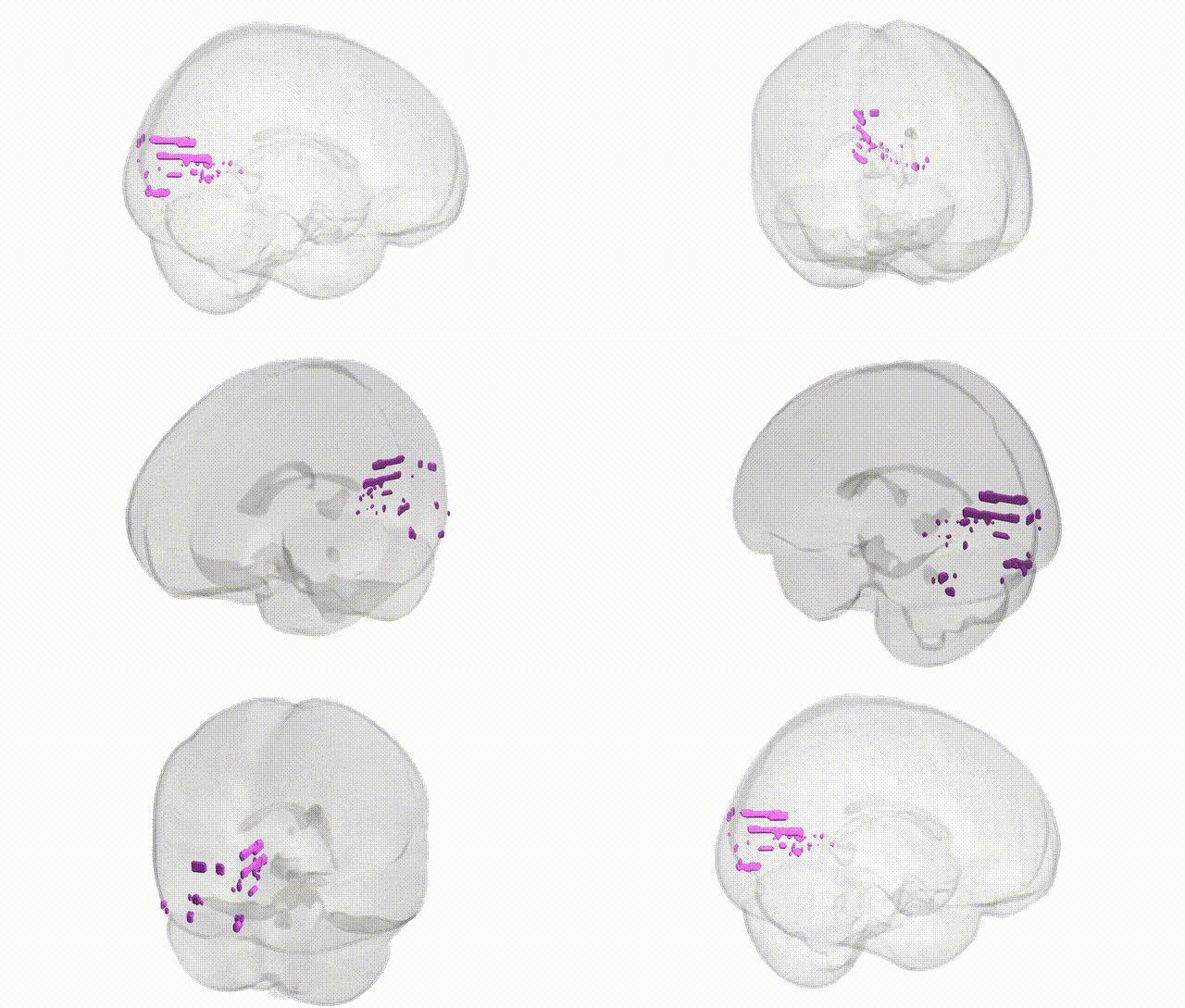
Full Quality Version: Download MP4
Wali Sidiqyar*, Gaurav Rudravaram*, Elyssa M. McMaster, Trent M. Schwartz, Adam M. Saunders, Kurt G. Schilling, Bennett A. Landman "Introducing SPINS: A Shared Public Visualization Library of Neuroanatomical Structures." Medical Imaging with Deep Learning- short paper
This resource is licensed under CC0 1.0 Universal (Public Domain).