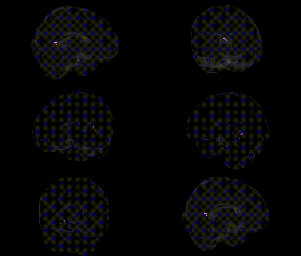
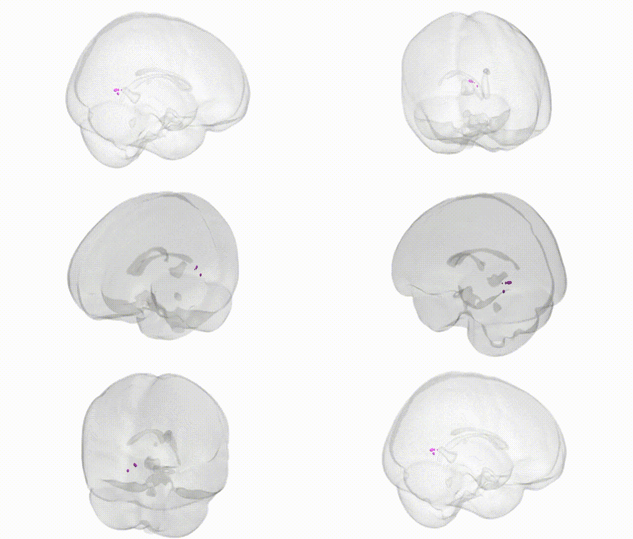
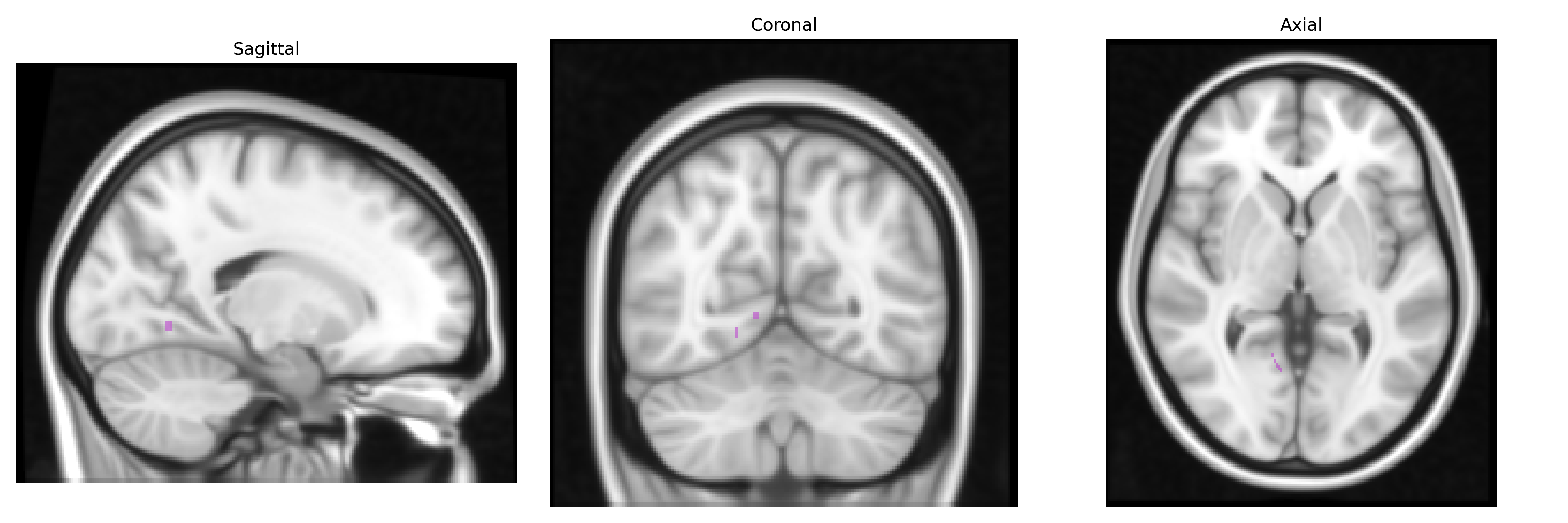
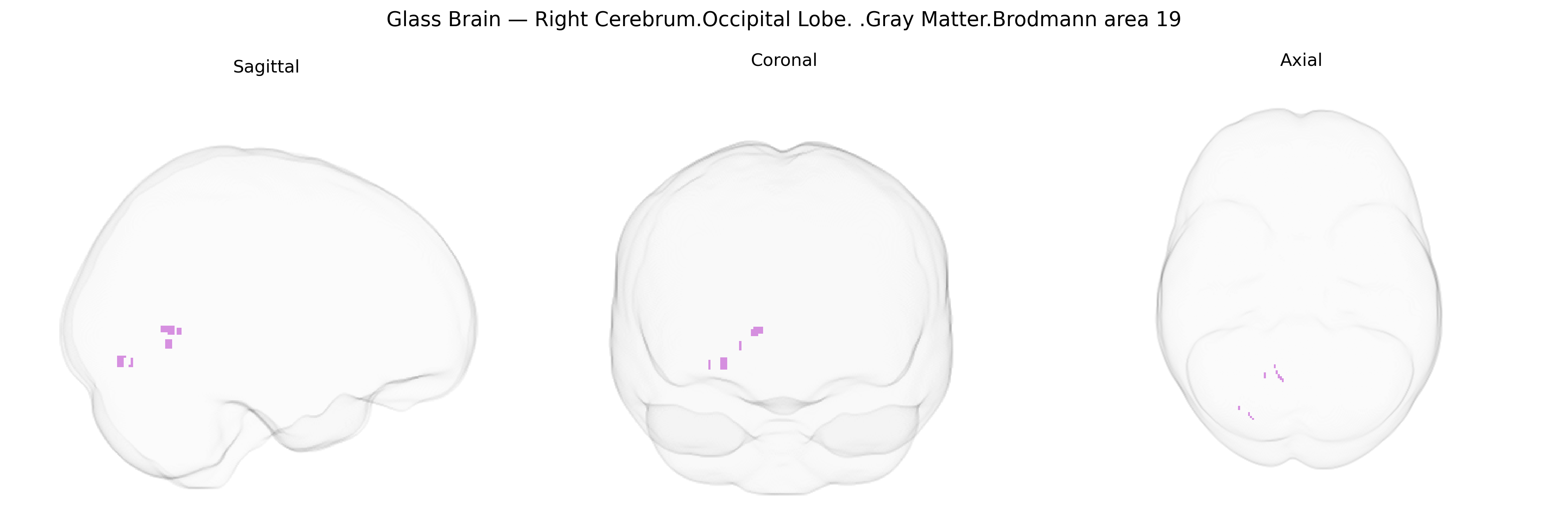
The bilateral Right Cerebrum.Occipital Lobe.Gray Matter.Brodmann area 19 corresponds to a higher-order visual association cortex located in the occipital lobe, bordering primary (BA17) and secondary (BA18) visual areas and extending into adjacent temporal and parietal regions. This region participates in complex visual processing tasks, including integration of motion, form, color, and spatial information, and contributes to object recognition, visual attention, and the interpretation of visual context. Neuronal populations here receive convergent input from earlier visual areas and relay processed signals to multimodal association cortices, supporting higher-level visual perception and visuocognitive functions. There is no direct link for Brodmann area 19; a related structure is Visual cortex.
Brodmann area 19 (BA19) in the bilateral right occipital lobe, a higher-order visual association region, has been implicated in several genetic and GWAS-based findings that primarily relate to visual processing, neurodevelopment, and psychiatric or cognitive traits. Large-scale imaging-genetics consortia (e.g., ENIGMA, UK Biobank) report that gray matter volume, cortical thickness, and surface area in occipital association cortices, including BA19, show significant heritability and associations with common variants in genes involved in neurodevelopment (such as MIR137, DISC1, and neuregulin/ErbB pathway genes), synaptic plasticity (e.g., BDNF, GRIN2B), and axon guidance (e.g., ROBO and semaphorin families). Occipital regions encompassing BA19 frequently emerge in GWAS of brain morphology and functional connectivity, which have linked variants in genes like HMGA2, KTN1, and others to global and regional cortical surface area, including visual association areas. Genetic studies of visual cortical plasticity and visual disorders, such as albinism, congenital blindness, and amblyopia, suggest that variants in melanogenesis and visual pathway genes (e.g., TYR, OCA2, GPR143) indirectly alter BA19 structure and function through atypical visual input, with downstream effects on gray matter volume and connectivity. Neuropsychiatric GWAS and imaging-genetics work have connected polygenic risk for schizophrenia, autism spectrum disorder, ADHD, and major depression with altered structure or activation in BA19 and neighboring occipital association cortex, implicating shared genetic architectures involving glutamatergic, GABAergic, and calcium-channel genes (e.g., CACNA1C). Additionally, GWAS of cognitive abilities and educational attainment identify polygenic influences that correlate with occipital association cortex characteristics, including BA19, suggesting that genetic variants affecting general cortical development, synaptic function, and white-matter organization contribute to individual differences in higher-order visual integration and visuospatial cognition mediated by this region.
Overview generated by GPT-4o (2026).
Region ID: 263
Hemisphere: bilateral
Atlas: Talairach labels 1mm
Full Quality Version: Download MP4
Full Quality Version: Download MP4
Wali Sidiqyar*, Gaurav Rudravaram*, Elyssa M. McMaster, Trent M. Schwartz, Adam M. Saunders, Kurt G. Schilling, Bennett A. Landman "Introducing SPINS: A Shared Public Visualization Library of Neuroanatomical Structures." Medical Imaging with Deep Learning- short paper
This resource is licensed under CC0 1.0 Universal (Public Domain).