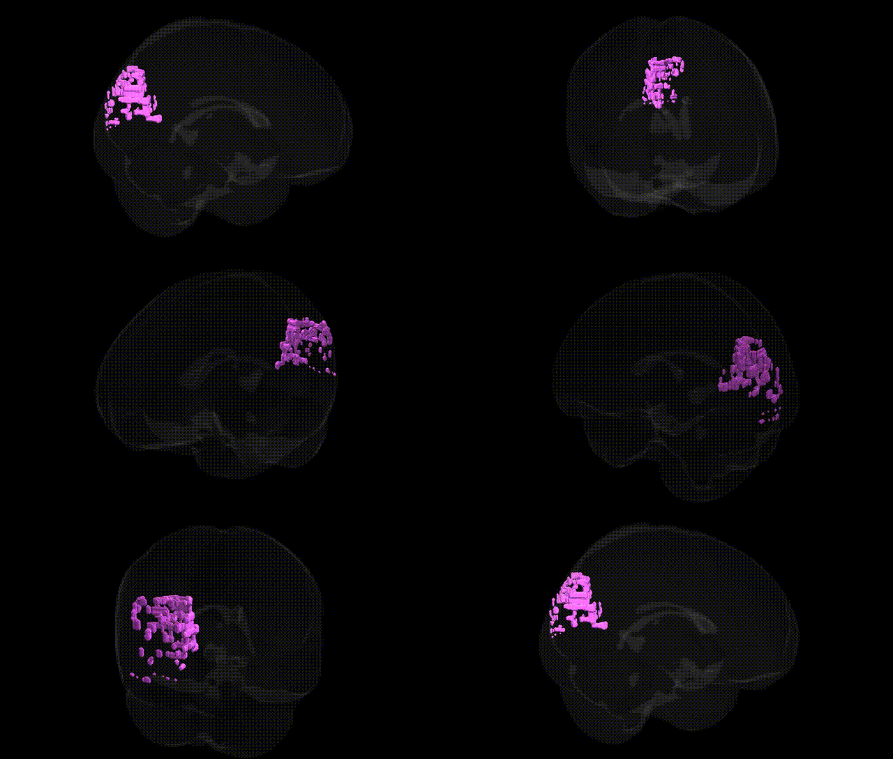
The bilateral Right Cerebrum.Occipital Lobe.Cuneus refers to the cuneus portion of the occipital cortex located in the dorsal medial aspect of the occipital lobe, bounded superiorly by the parieto-occipital sulcus and inferiorly by the calcarine sulcus. This region is part of the primary and associative visual cortices and is crucial for basic visual processing, particularly involving visual attention, spatial orientation, and aspects of visual pattern recognition. It receives input from the lateral geniculate nucleus via the optic radiations and participates in processing information related to the central visual field, integrating signals that contribute to conscious visual perception and visually guided behavior. Cuneus
The bilateral right cuneus (Right Cerebrum.Occipital Lobe.Cuneus) has been implicated in several genetic and GWAS-based associations, primarily through its role in visual processing and large-scale brain-structure and connectivity phenotypes. Numerous imaging-genetics studies (e.g., UK Biobank–based GWAS of cortical thickness, surface area, and functional connectivity) have identified common variants in genes involved in neurodevelopment, synaptic function, and myelination—such as MIR137, GRIN2B, CACNA1C, and genes in the 22q11.2 locus—that show effects on occipital and cuneus morphology or activity, often in the context of schizophrenia, bipolar disorder, and major depression. Polygenic risk scores for schizophrenia, autism spectrum disorder, and ADHD have been associated with altered cuneus thickness, surface area, or functional connectivity, suggesting that visual and higher-order integration areas are sensitive to neurodevelopmental risk. GWAS of resting-state networks and visual cortex activation have identified associations with loci related to neuronal differentiation (e.g., ROBO and semaphorin pathways) and with genes influencing intracranial volume and overall cortical architecture (e.g., HMGA2, IGF1-related pathways), some of which show specific or stronger effects in occipital regions, including the cuneus. Additionally, rare variant and CNV studies in disorders with visual or visuospatial dysfunction (e.g., certain forms of epilepsy, neurodegenerative disease, and congenital visual pathway anomalies) have occasionally reported structural or functional cuneus involvement, although these findings are usually secondary to broader posterior cortical or occipital lobe abnormalities rather than uniquely localized to the right cuneus.
Overview generated by GPT-4o (2026).
Region ID: 476
Hemisphere: bilateral
Atlas: Talairach labels 1mm
Full Quality Version: Download MP4
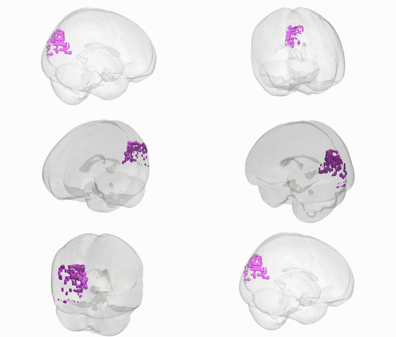
Full Quality Version: Download MP4
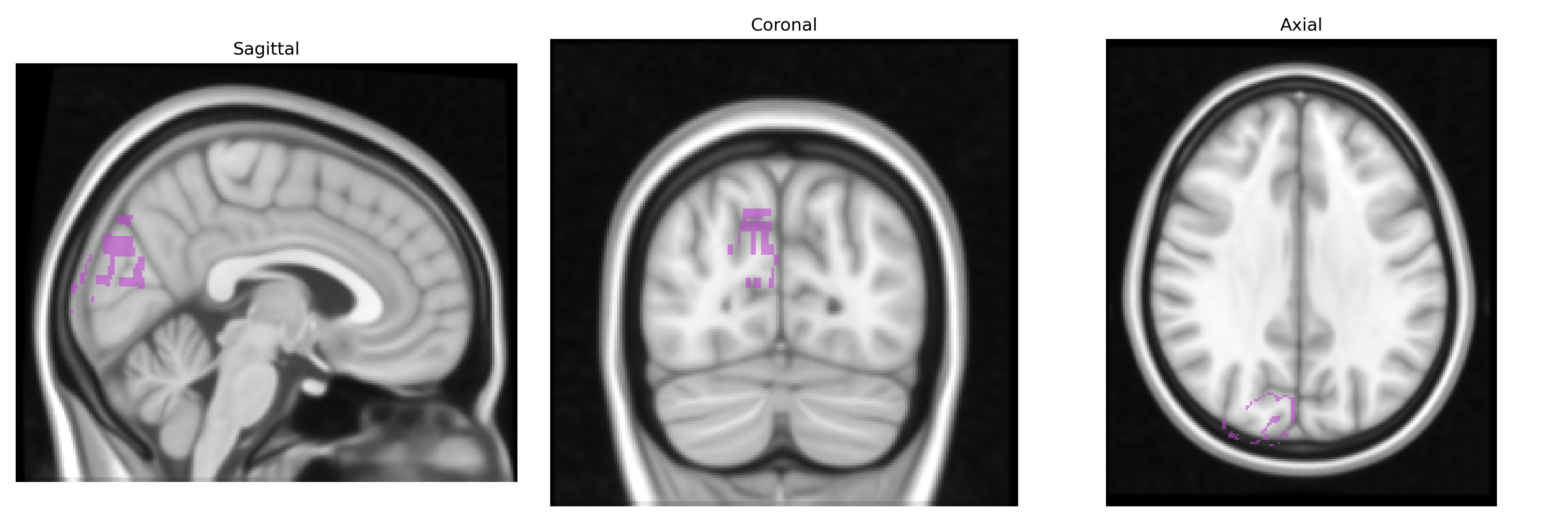
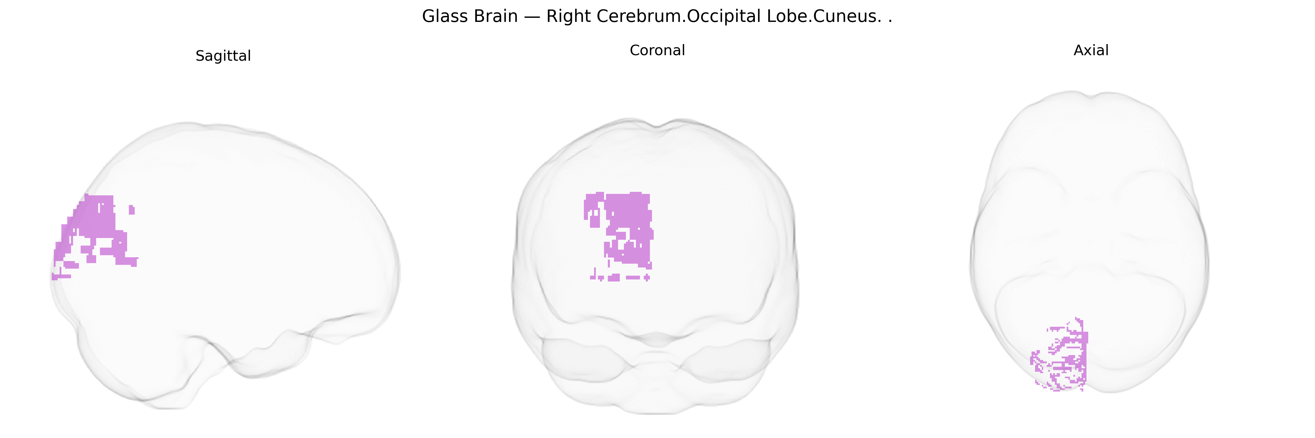
Wali Sidiqyar*, Gaurav Rudravaram*, Elyssa M. McMaster, Trent M. Schwartz, Adam M. Saunders, Kurt G. Schilling, Bennett A. Landman "Introducing SPINS: A Shared Public Visualization Library of Neuroanatomical Structures." Medical Imaging with Deep Learning- short paper
This resource is licensed under CC0 1.0 Universal (Public Domain).