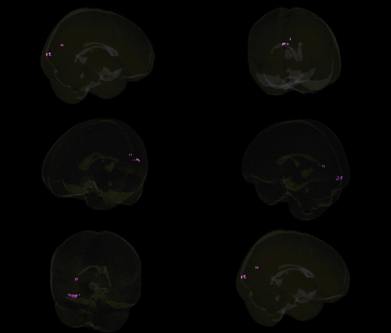
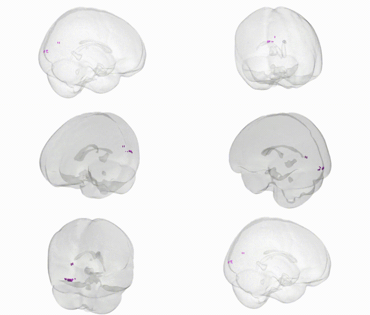
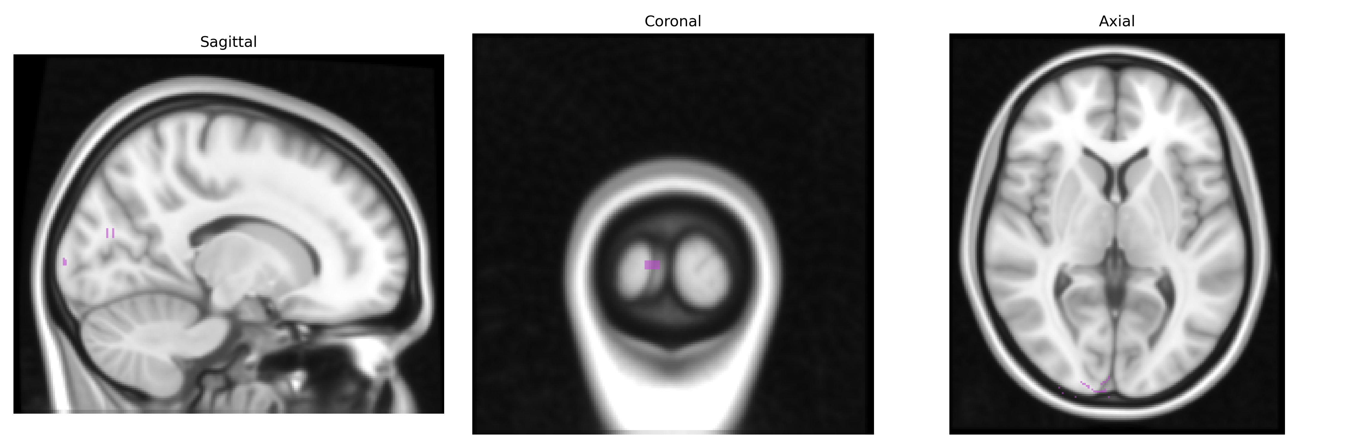
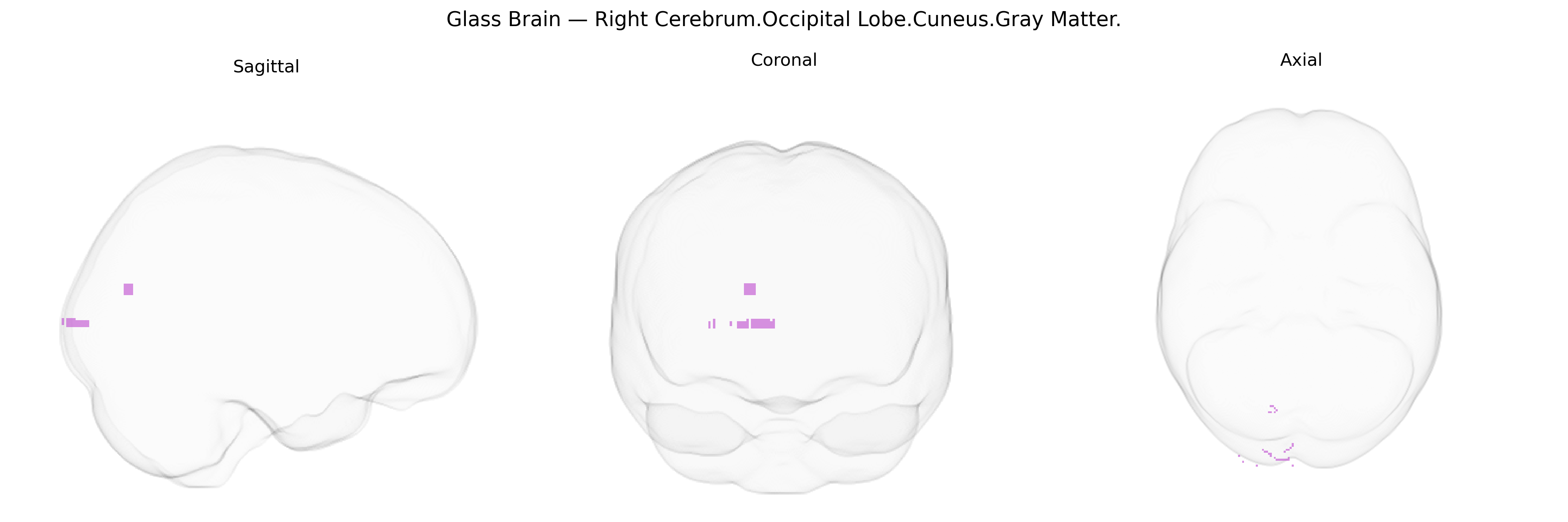
The bilateral Right Cerebrum.Occipital Lobe.Cuneus.Gray Matter corresponds to the cortical gray matter of the cuneus, a wedge-shaped region of the occipital lobe situated on the medial surface of the hemisphere between the parieto‑occipital sulcus and the calcarine sulcus. This area forms part of the primary and secondary visual cortices, receiving and processing retinotopically organized visual input, particularly from the central visual field, and contributes to basic visual perception, including orientation, spatial processing, and integration of visual signals for higher-order interpretation. The cuneus participates in visual attention, visual imagery, and aspects of visuospatial cognition, and is interconnected with other occipital, parietal, and temporal visual association areas as well as subcortical visual relay nuclei. Cuneus
The cuneus gray matter of the occipital lobe, as defined in the Talairach 1 mm atlas, shows genetic associations primarily related to visual processing, cortical thickness, and broader neuropsychiatric risk, rather than a single specific disorder; large-scale imaging–genetics consortia (e.g., ENIGMA, UK Biobank) have identified common variants in genes involved in neurodevelopment, synaptic function, and myelination (such as loci near MAPT, HMGA2, and multiple 3p, 6q, and 17q regions) that influence cuneus cortical thickness or surface area, and these structural endophenotypes partially mediate polygenic risk for disorders including schizophrenia, major depression, and bipolar disorder. GWAS of brain morphology and functional connectivity frequently implicate the cuneus as part of visual and default mode networks, with overlap between variants affecting cuneus volume and those associated with cognitive performance, educational attainment, and general intelligence, suggesting shared genetic architecture between occipital structural variation and higher-order cognition. Additional associations link cuneus measures to migraine and visual aura susceptibility, as well as to neurodegenerative traits where occipital atrophy features (e.g., posterior cortical atrophy and some Alzheimer’s disease endophenotypes), indicating that genetic variants influencing this region may contribute to individual differences in visual integration, vulnerability to visual–cognitive syndromes, and broad neuropsychiatric liability.
Overview generated by GPT-4o (2026).
Region ID: 535
Hemisphere: bilateral
Atlas: Talairach labels 1mm
Full Quality Version: Download MP4
Full Quality Version: Download MP4
Wali Sidiqyar*, Gaurav Rudravaram*, Elyssa M. McMaster, Trent M. Schwartz, Adam M. Saunders, Kurt G. Schilling, Bennett A. Landman "Introducing SPINS: A Shared Public Visualization Library of Neuroanatomical Structures." Medical Imaging with Deep Learning- short paper
This resource is licensed under CC0 1.0 Universal (Public Domain).