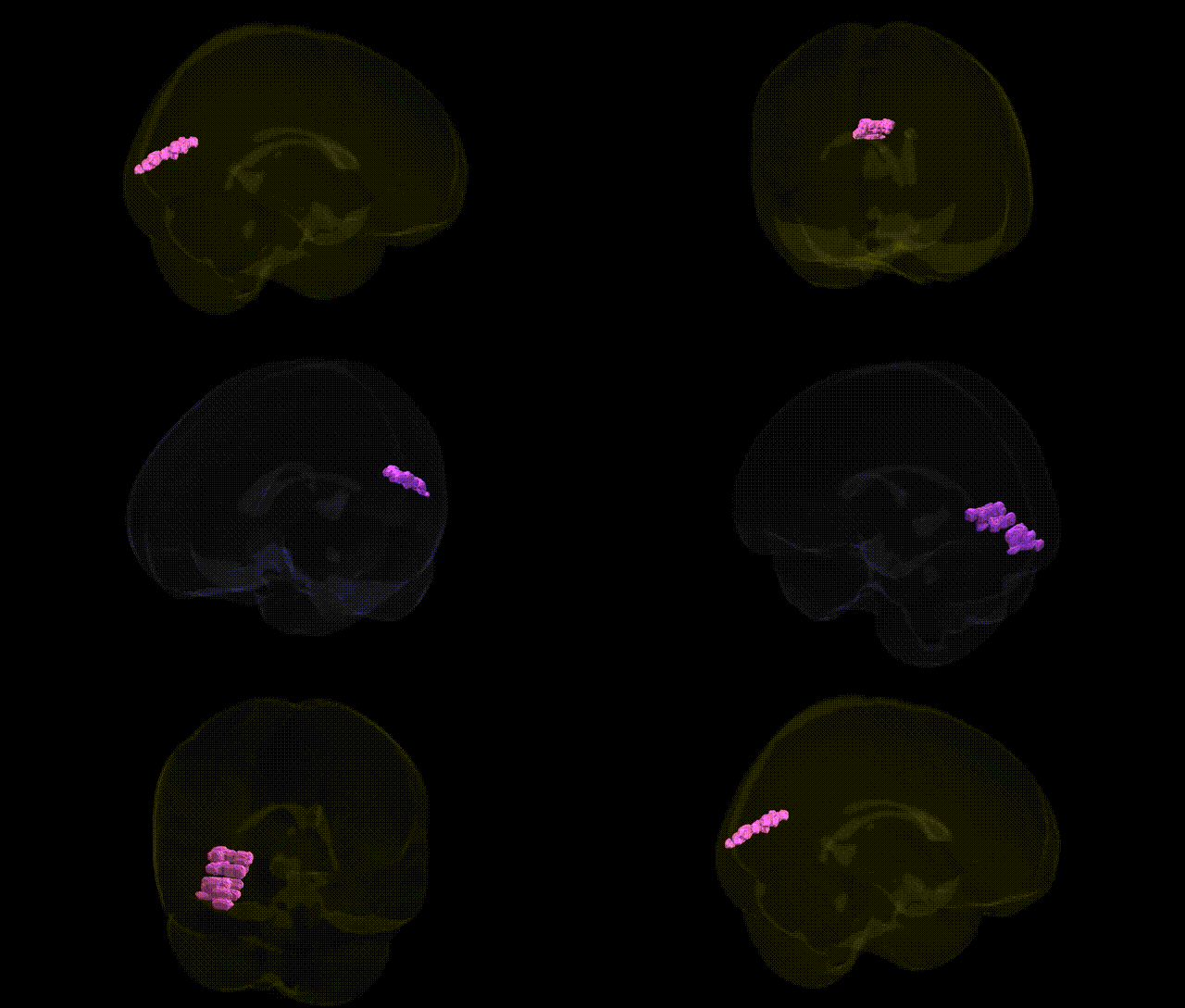
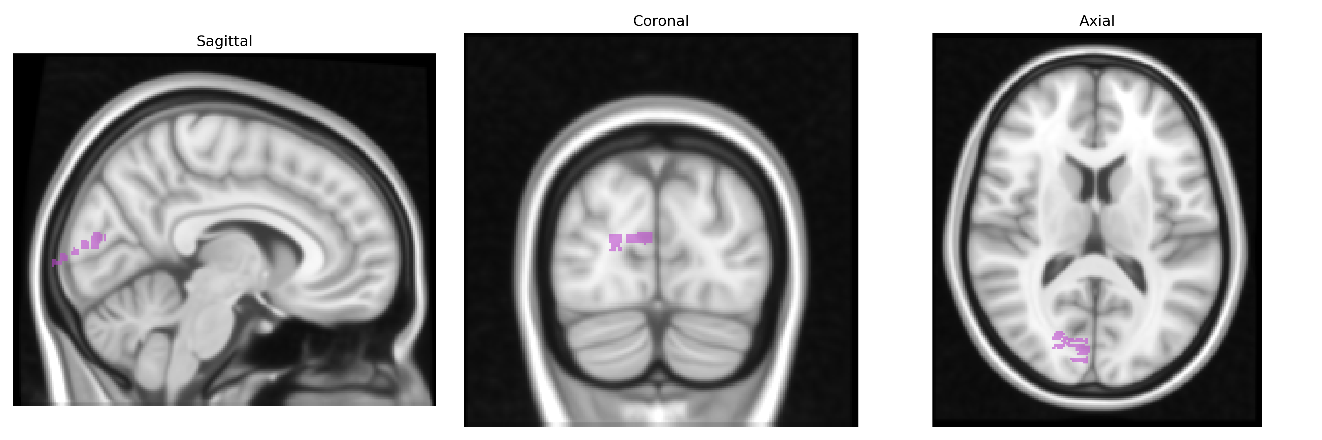
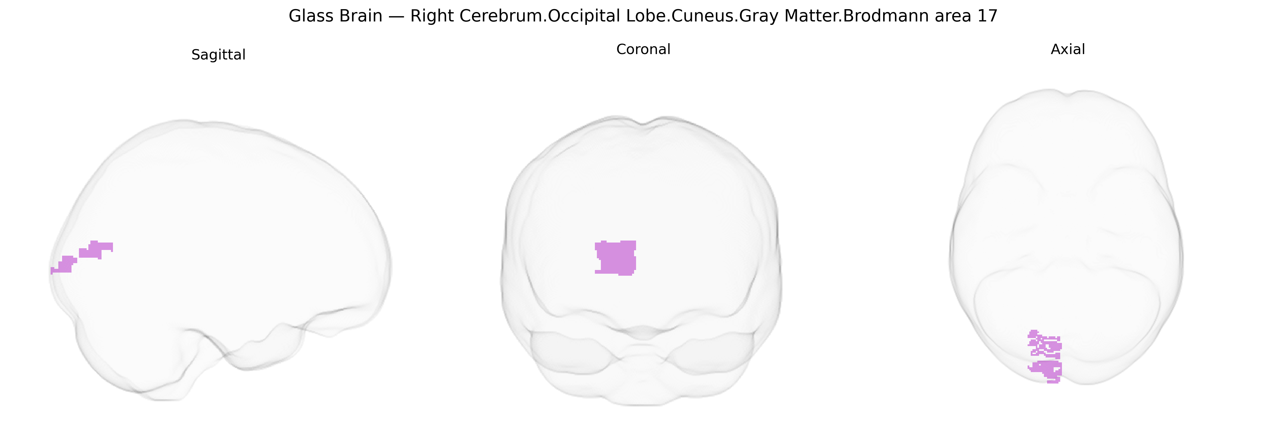
The bilateral Right Cerebrum.Occipital Lobe.Cuneus.Gray Matter.Brodmann area 17 corresponds to the primary visual cortex (V1) located along the calcarine fissure in the medial aspect of the occipital lobe. This cytoarchitectonically defined region receives the densest input from the lateral geniculate nucleus of the thalamus and is organized retinotopically, with distinct portions representing specific regions of the visual field. Neurons in this area are specialized for processing fundamental visual features such as orientation, spatial frequency, contrast, and motion direction, forming the initial cortical stage of visual perception. Its laminar structure is highly differentiated, especially the prominent layer IV, which is subdivided into multiple sublayers corresponding to magnocellular and parvocellular inputs. Functionally, this region is essential for conscious visual experience and serves as the principal gateway to higher-order visual association areas in the occipital and temporal lobes.
Primary visual cortex
The bilateral right occipital lobe cuneus gray matter Brodmann area 17 (primary visual cortex, V1) has been implicated in genetic studies largely through its role in visual processing and structural variation. Twin and imaging-genetics studies show high heritability of V1 surface area and cortical thickness, with polygenic influences distributed across the genome rather than single major loci. GWAS of occipital and V1 cortical measures (e.g., ENIGMA and UK Biobank cohorts) have identified common variants near genes involved in neurodevelopment and synaptic function, including regulatory loci influencing cortical surface area and thickness, although specific, robust BA17-exclusive associations are limited. Genetic variants in visual system genes such as RGS4, GRIN2B, and others have been linked to functional activation differences in occipital cortex during visual tasks, and polymorphisms in serotonin and dopamine-related genes have been associated with altered visual cortex responses in fMRI paradigms. Occipital and BA17 abnormalities have been reported in genetically influenced disorders such as schizophrenia, bipolar disorder, major depression, and autism spectrum disorder, with GWAS-implicated risk genes (e.g., complement pathway genes, calcium channel genes like CACNA1C, and synaptic genes such as NRXN1) contributing to altered visual processing and structural changes in this region, though these associations reflect broad brain-wide effects rather than specificity to BA17. In migraine with aura, rare variants and GWAS signals affecting cortical excitability and visual pathways have been linked to altered V1 responsivity, and in dyslexia and other reading or visual-perceptual disorders, common variants in neurodevelopmental genes show associations with occipital and cuneus activation and morphology. Overall, genetic influences on BA17 are robust at the level of heritable structure and function, but current GWAS and imaging-genetic findings point to polygenic, distributed effects with limited evidence for region-specific genetic loci unique to this Talairach-defined area.
Overview generated by GPT-4o (2026).
Region ID: 477
Hemisphere: bilateral
Atlas: Talairach labels 1mm
Full Quality Version: Download MP4
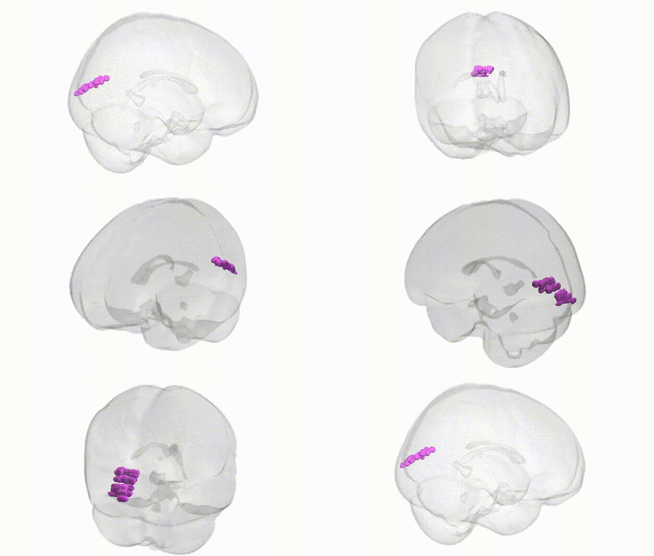
Full Quality Version: Download MP4
Wali Sidiqyar*, Gaurav Rudravaram*, Elyssa M. McMaster, Trent M. Schwartz, Adam M. Saunders, Kurt G. Schilling, Bennett A. Landman "Introducing SPINS: A Shared Public Visualization Library of Neuroanatomical Structures." Medical Imaging with Deep Learning- short paper
This resource is licensed under CC0 1.0 Universal (Public Domain).