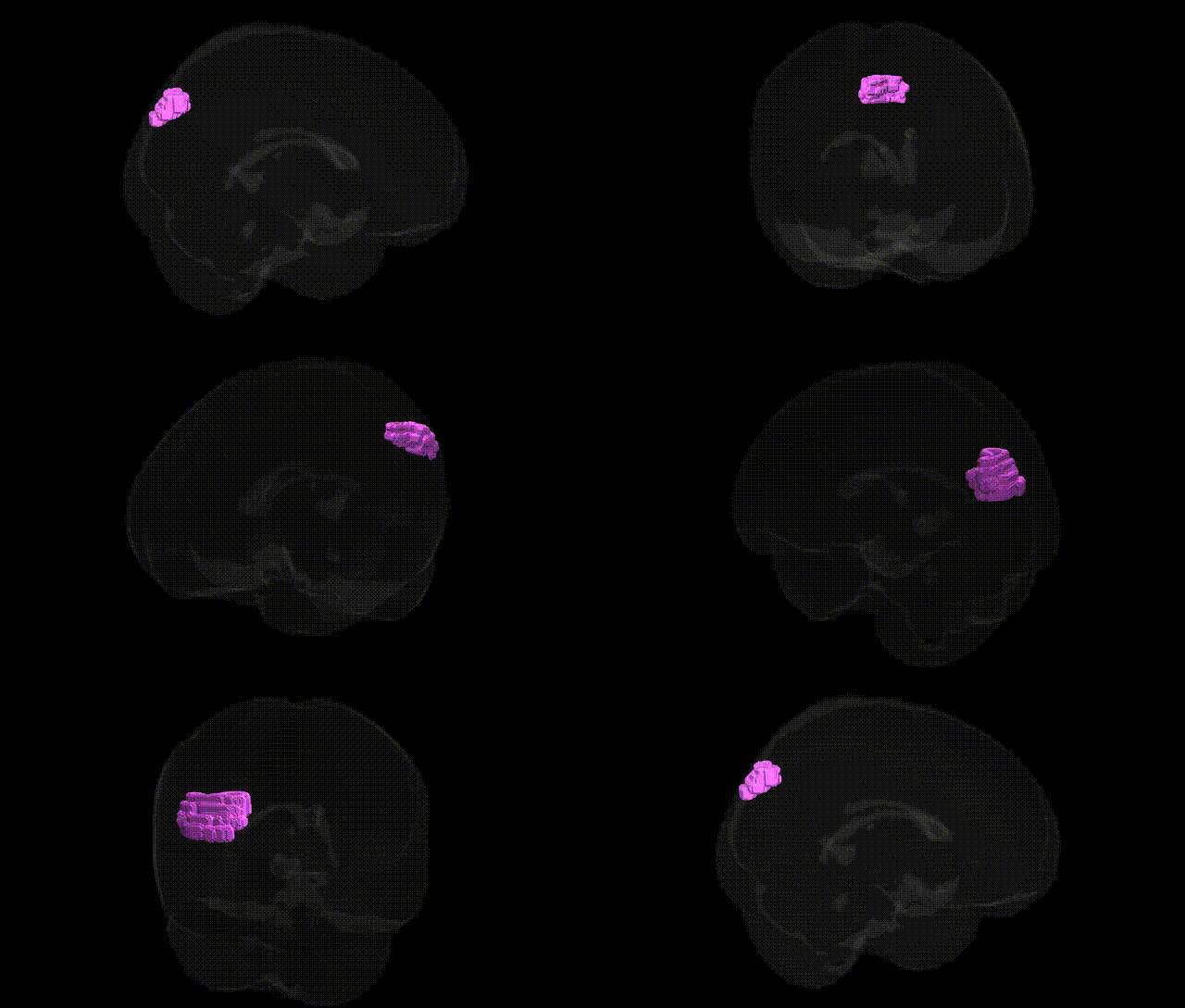
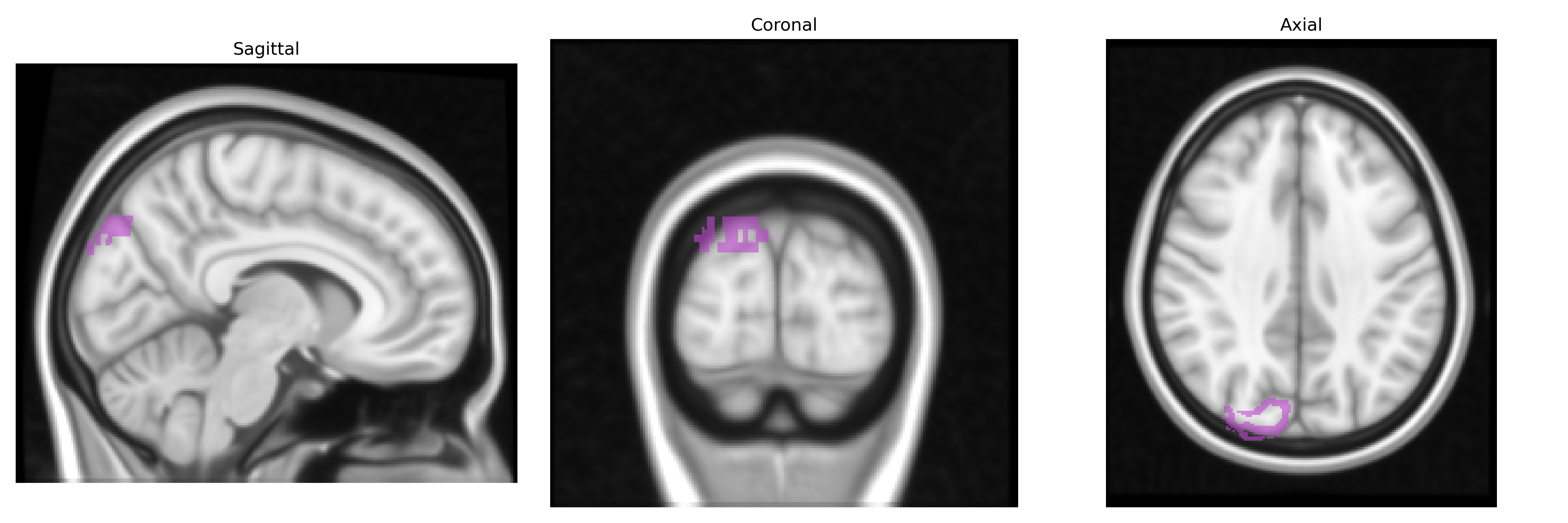
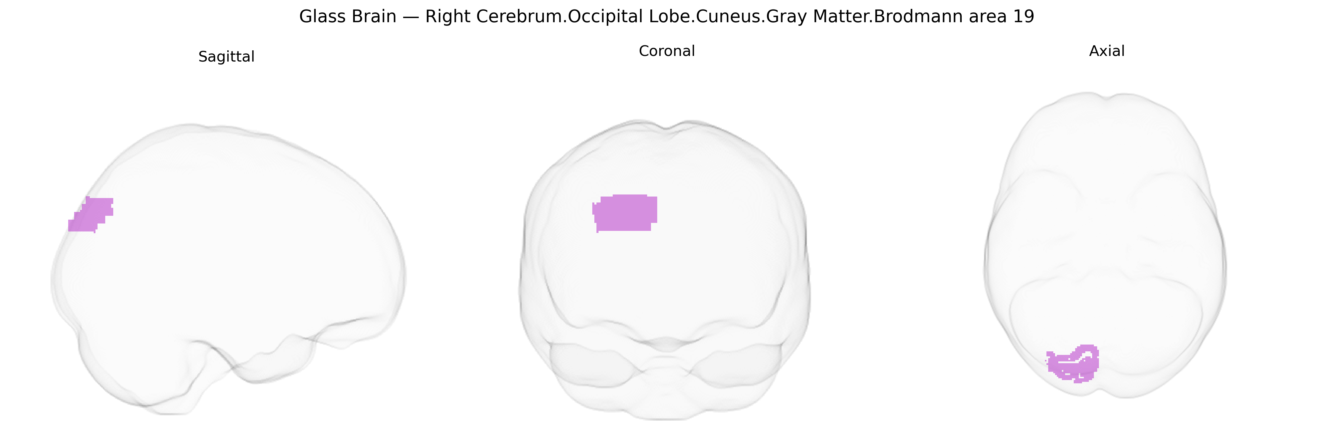
The bilateral Right Cerebrum.Occipital Lobe.Cuneus.Gray Matter.Brodmann area 19 corresponds to a higher-order visual associative cortical region located in the dorsal occipital lobe, within the cuneus of the right hemisphere, and extending medially along the calcarine fissure posterior to primary visual cortex (BA17) and contiguous with secondary visual cortex (BA18). Histologically, Brodmann area 19 exhibits a well-developed granular layer IV and dense myelination patterns characteristic of extrastriate visual areas, integrating complex visual inputs for functions such as motion processing, shape and form analysis, and aspects of visuospatial perception, including contributions to object recognition and the integration of visual information with higher-order association cortices. There is no direct link for this exact composite Talairach label; a closely related structure is Brodmann area 19.
The bilateral Right Cerebrum.Occipital Lobe.Cuneus.Gray Matter.Brodmann area 19, a higher-order visual association region, has been implicated in several genetic and GWAS-based findings linking structural or functional variation to neuropsychiatric, neurodevelopmental, and visual traits. Twin and family studies indicate substantial heritability of gray matter volume and surface area in BA19/cuneus, with contributions from common variants in genes involved in synaptic plasticity, neurodevelopment, and myelination (e.g., BDNF, NRG1, and genes in glutamatergic and GABAergic pathways), although locus-level specificity is often shared with other occipital and parietal regions. Large-scale imaging-genetics consortia (e.g., ENIGMA, UK Biobank) have identified associations between occipital/cuneus cortical thickness or volume and variants near genes involved in axon guidance and neuronal patterning (such as DCC, ROBO family members, and protocadherins), as well as polygenic scores for cognitive ability and educational attainment, suggesting that BA19 morphology reflects distributed genetic influences on visual–cognitive processing. Functional imaging–genetics work links variation in dopaminergic and serotonergic genes (e.g., COMT, 5-HTTLPR) to BA19 activation differences during visual attention and emotional face processing tasks, implicating this region in genetically modulated visual–affective integration. Clinically, BA19/cuneus alterations have been associated with genetic risk for schizophrenia, major depressive disorder, and autism spectrum conditions, where risk loci and polygenic risk scores correlate with reduced occipital gray matter or altered connectivity involving BA19; additional associations are reported with migraine, photosensitivity-related traits, and visual hallucinations in disorders such as Parkinson’s disease, though these effects typically arise from broader visual network and neurotransmission genes rather than BA19-specific loci.
Overview generated by GPT-4o (2026).
Region ID: 844
Hemisphere: bilateral
Atlas: Talairach labels 1mm
Full Quality Version: Download MP4
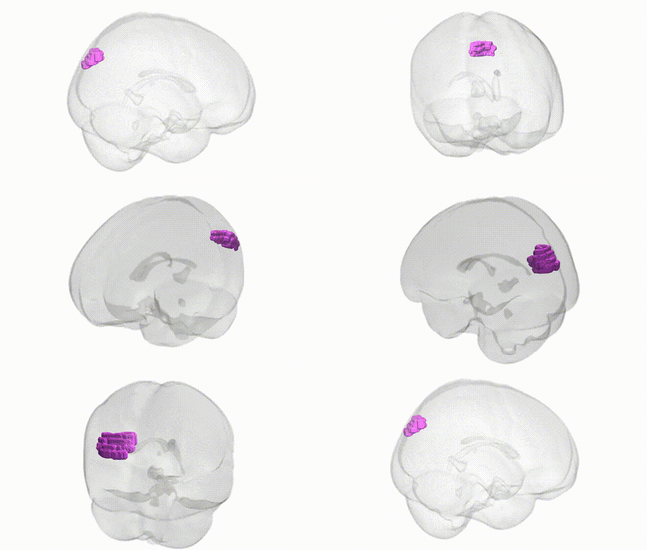
Full Quality Version: Download MP4
Wali Sidiqyar*, Gaurav Rudravaram*, Elyssa M. McMaster, Trent M. Schwartz, Adam M. Saunders, Kurt G. Schilling, Bennett A. Landman "Introducing SPINS: A Shared Public Visualization Library of Neuroanatomical Structures." Medical Imaging with Deep Learning- short paper
This resource is licensed under CC0 1.0 Universal (Public Domain).