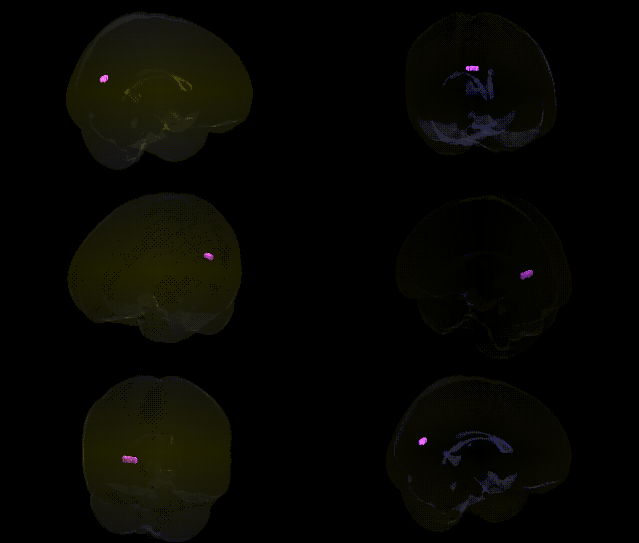
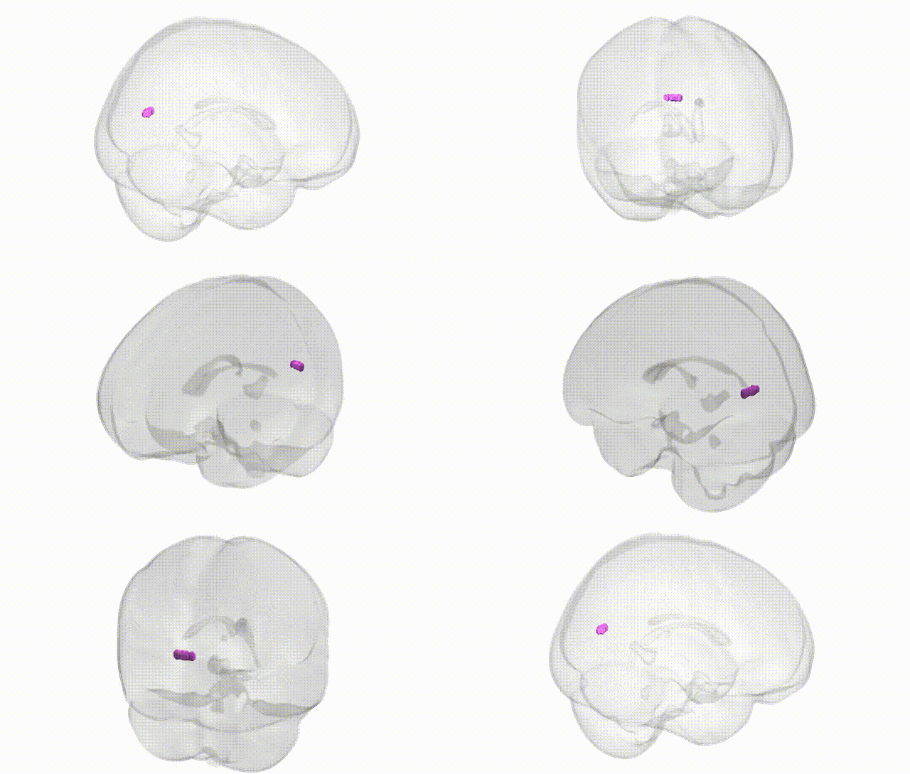
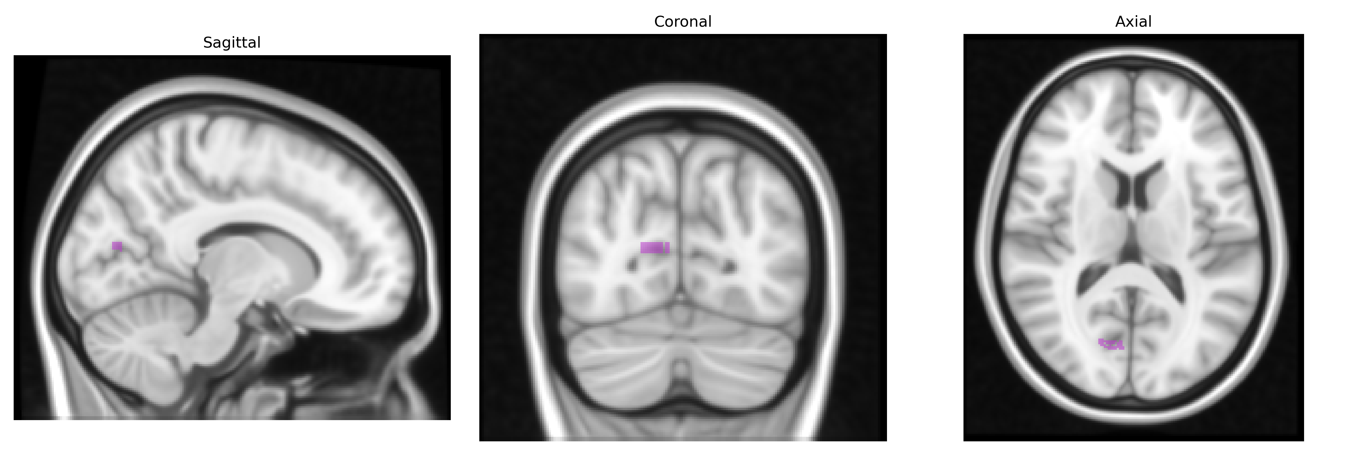
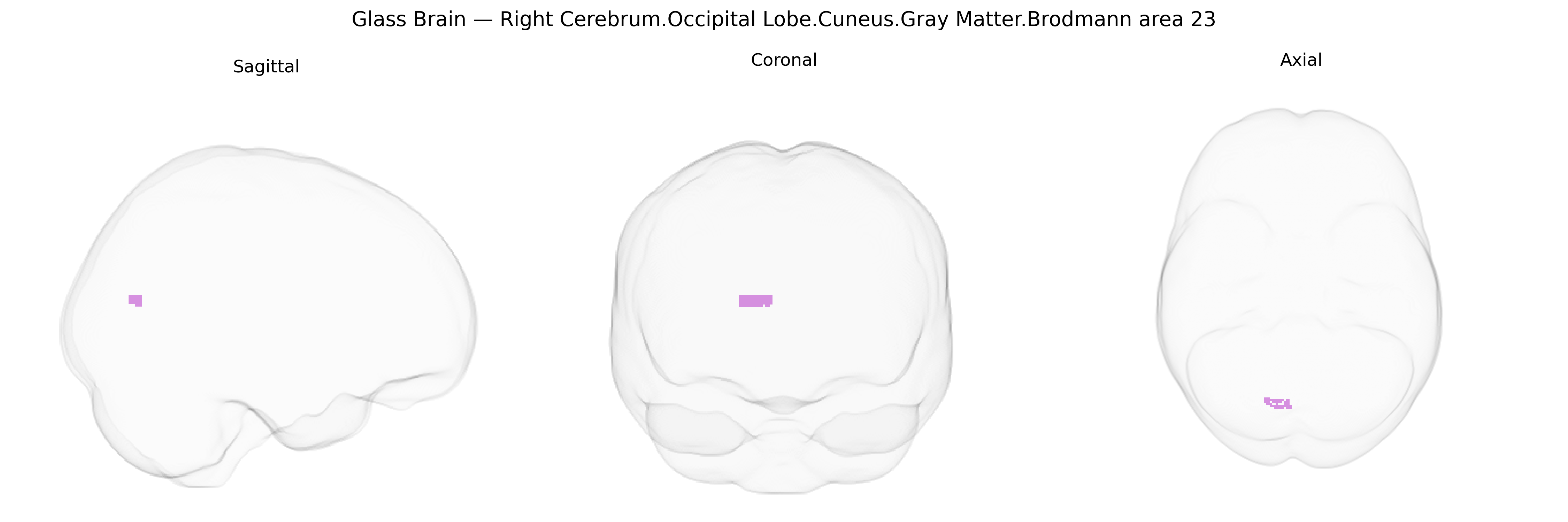
The bilateral Right Cerebrum.Occipital Lobe.Cuneus.Gray Matter.Brodmann area 23 corresponds to gray matter within the cuneus region of the occipital lobe that has been functionally or cytoarchitectonically associated with Brodmann area 23, a subdivision of the posterior cingulate cortex in the limbic lobe. In the Talairach 1 mm atlas, this labeling reflects a combination of anatomical (cuneus, occipital lobe) and Brodmann area–based parcellation, capturing a region implicated in higher-order visual processing, visuospatial integration, and interactions between visual input and limbic or associative networks. Although Brodmann area 23 itself is classically described in the posterior cingulate cortex rather than in the cuneus, atlas-based extensions can map BA23 labels into adjacent medial occipital territory, particularly where retrosplenial and posterior cingulate areas border visual cortex. This region participates in networks subserving visual context evaluation, memory-related scene processing, and integration of visual information with internal states and self-referential processing. (No direct article exists for this exact composite label; a related structure is Brodmann area 23.)
The bilateral right cuneus gray matter in the occipital lobe (Talairach 1 mm, Brodmann area 23 labeling here reflects a likely parcellation/atlas mismatch, as BA23 is classically retrosplenial/posterior cingulate rather than occipital) has been implicated in several genetic and GWAS-based associations through imaging-genetics and neuropsychiatric studies, although few findings are specific to this exact micro-anatomical label. Large-scale ENIGMA and UK Biobank imaging GWAS have linked variants in genes involved in neurodevelopment, synaptic function, and myelination (e.g., HMGA2, IGF1, MAPT-region loci, and multiple chromatin-remodeling genes) to occipital and cuneus cortical thickness, surface area, and gray-matter volume, traits that often encompass this region. These structural and functional metrics in the cuneus/medial occipital cortex have shown genetic correlations and shared loci with disorders such as major depressive disorder, schizophrenia, autism spectrum disorder, and Alzheimer’s disease, as well as with cognitive traits (general intelligence, visual processing speed) and neuroticism, though effects are typically modest and distributed across widespread networks. Polygenic risk for schizophrenia and depression has been associated with altered cuneus/occipital activation in fMRI tasks and resting-state connectivity, particularly within default mode and visual networks, and APOE ε4 and other Alzheimer’s disease risk variants have been linked to early changes in medial occipital/posterior cingulate regions that sometimes anatomically overlap the atlas-defined area. Overall, current evidence supports a polygenic architecture in which numerous common variants contribute to structural and functional variability in this region, but no single gene or variant is uniquely or specifically tied to the bilateral right cuneus/BA23 label.
Overview generated by GPT-4o (2026).
Region ID: 707
Hemisphere: bilateral
Atlas: Talairach labels 1mm
Full Quality Version: Download MP4
Full Quality Version: Download MP4
Wali Sidiqyar*, Gaurav Rudravaram*, Elyssa M. McMaster, Trent M. Schwartz, Adam M. Saunders, Kurt G. Schilling, Bennett A. Landman "Introducing SPINS: A Shared Public Visualization Library of Neuroanatomical Structures." Medical Imaging with Deep Learning- short paper
This resource is licensed under CC0 1.0 Universal (Public Domain).