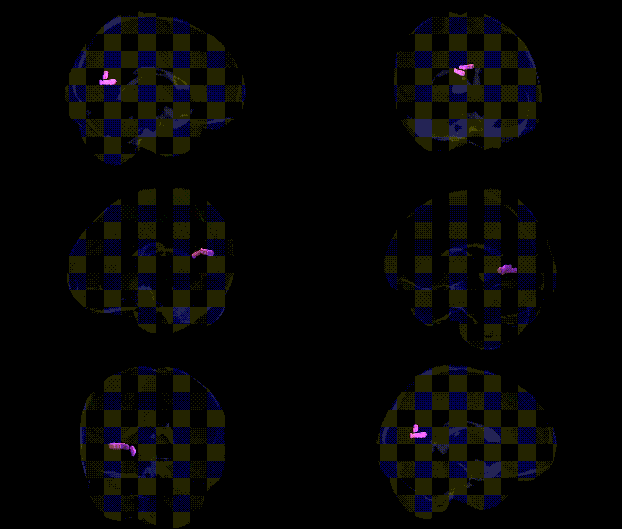
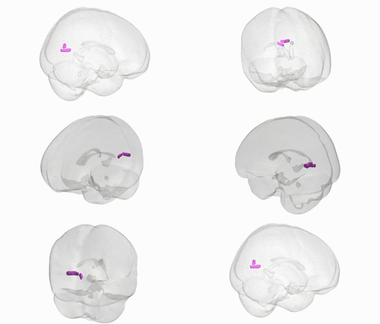
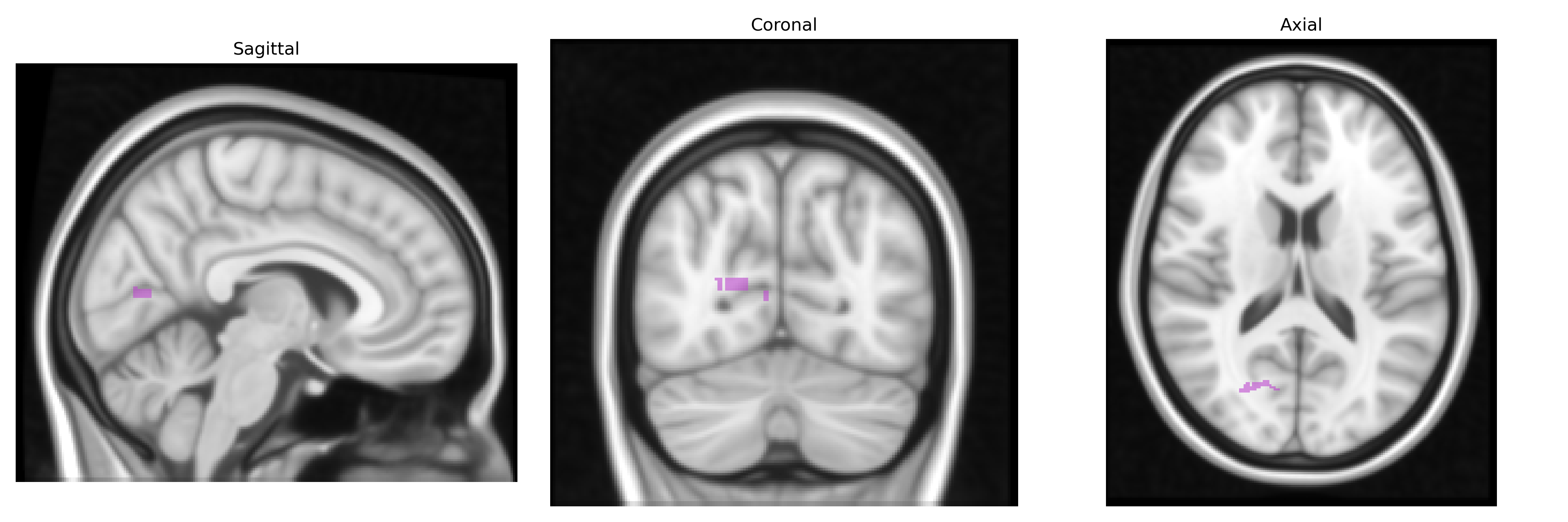
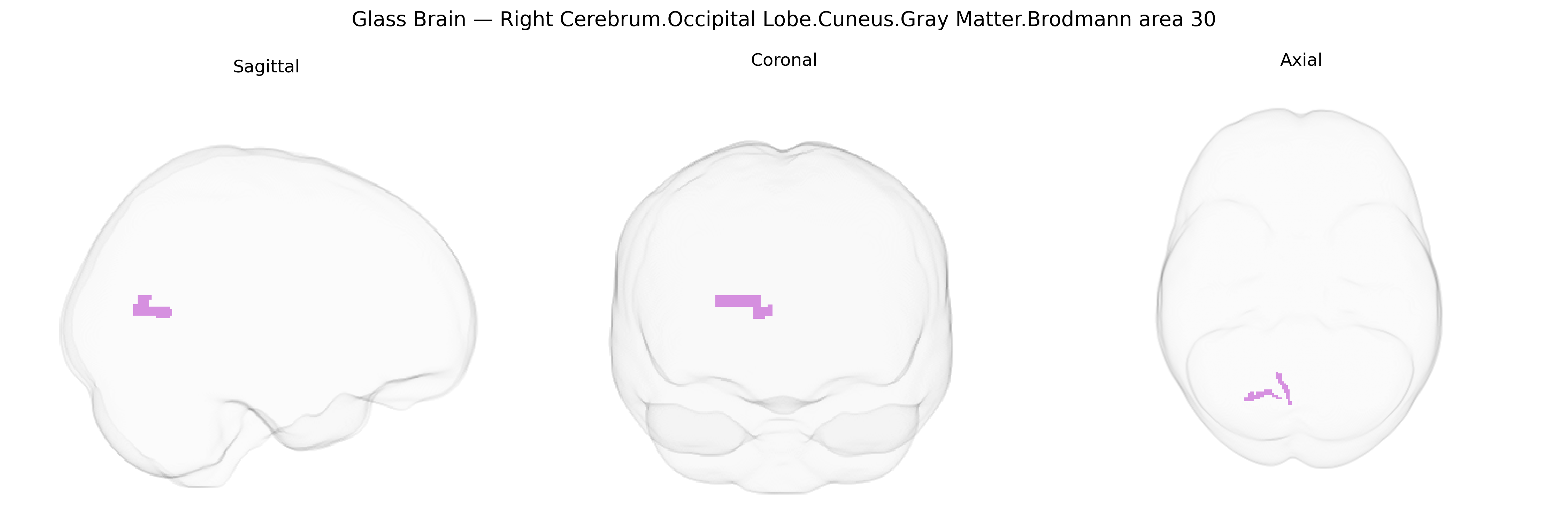
The bilateral Right Cerebrum.Occipital Lobe.Cuneus.Gray Matter.Brodmann area 30 corresponds to a retrosplenial portion of the cuneus within the medial occipital cortex, forming part of the ventral posterior cingulate/retrosplenial region involved in visuospatial processing, scene recognition, memory, and navigation. Brodmann area 30 lies adjacent to other retrosplenial and medial parietal areas (e.g., BA 23, 29, 31) and is situated near the parieto-occipital sulcus, integrating visual information with limbic and mnemonic networks. Functionally, this cortex participates in contextual and episodic memory, contributes to the default mode network, and shows strong connectivity with hippocampal–parahippocampal and posterior cingulate structures. There is no direct link for Brodmann area 30; a closely related structure is the Retrosplenial cortex.
The bilateral right cuneus gray matter in Brodmann area 30, a retrosplenial/occipital region, has been implicated in genetic studies largely through imaging genetics and GWAS of brain structure and function, rather than region-specific gene discoveries. Large-scale MRI-based GWAS (e.g., ENIGMA, UK Biobank) have identified multiple common variants associated with occipital and cuneus cortical thickness, surface area, and volume, frequently implicating genes involved in neurodevelopmental and synaptic pathways (such as those near MEF2C, TCF4, and other regulators of cortical patterning), though these findings usually refer to broader occipital or visual association areas rather than BA30 alone. Functionally, BA30 participates in visuospatial processing, scene memory, and navigation, and genetic variation in this region’s structure or activation has been linked to cognitive traits including general intelligence, memory performance, and educational attainment in polygenic score and imaging-genetics analyses. The cuneus/retrosplenial region also shows altered morphology and connectivity in psychiatric disorders—particularly major depressive disorder, schizophrenia, and bipolar disorder—where GWAS-implicated risk genes (e.g., CACNA1C, GRIN2A, and others involved in glutamatergic and calcium signaling) are thought to influence network-level changes that include BA30, though associations remain indirect and network-based rather than specific to this Brodmann area. Additionally, occipital and cuneus measures have modest genetic correlations with migraine, visual hallucinations in psychosis, and neurodegenerative conditions such as Alzheimer’s disease, suggesting shared polygenic influences on visual and memory-related circuitry, but no single gene or variant has been conclusively established as uniquely linked to the right BA30 cuneus in current GWAS catalogs.
Overview generated by GPT-4o (2026).
Region ID: 661
Hemisphere: bilateral
Atlas: Talairach labels 1mm
Full Quality Version: Download MP4
Full Quality Version: Download MP4
Wali Sidiqyar*, Gaurav Rudravaram*, Elyssa M. McMaster, Trent M. Schwartz, Adam M. Saunders, Kurt G. Schilling, Bennett A. Landman "Introducing SPINS: A Shared Public Visualization Library of Neuroanatomical Structures." Medical Imaging with Deep Learning- short paper
This resource is licensed under CC0 1.0 Universal (Public Domain).