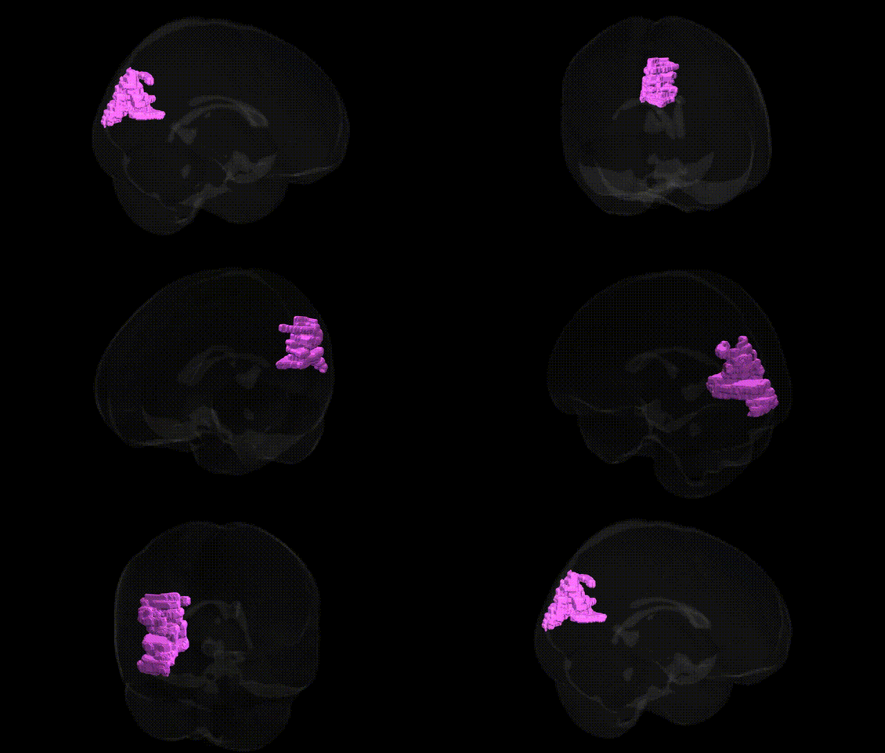
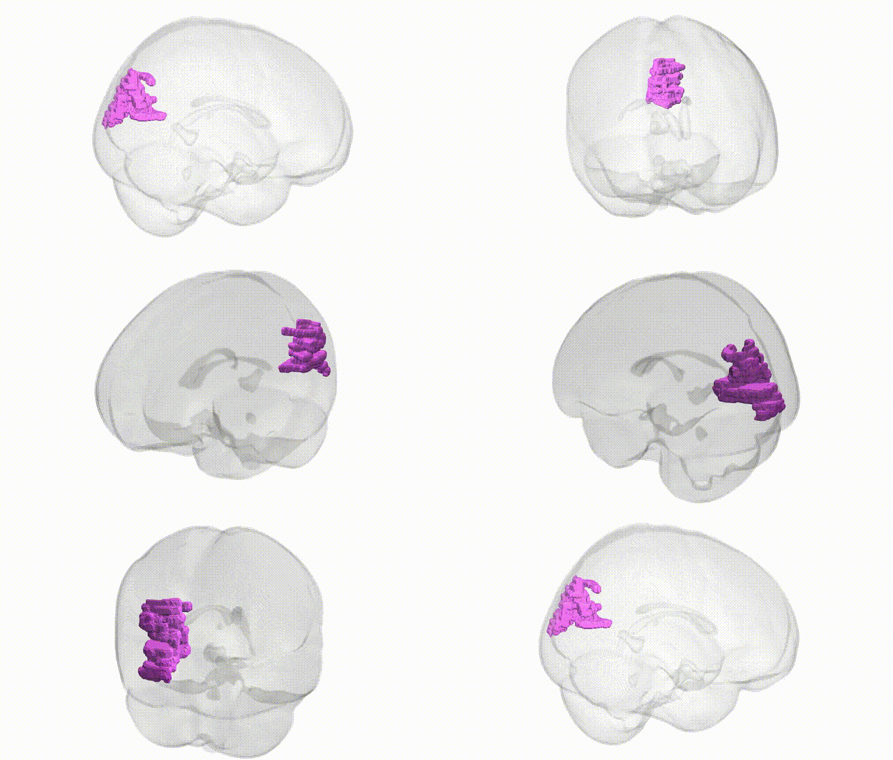
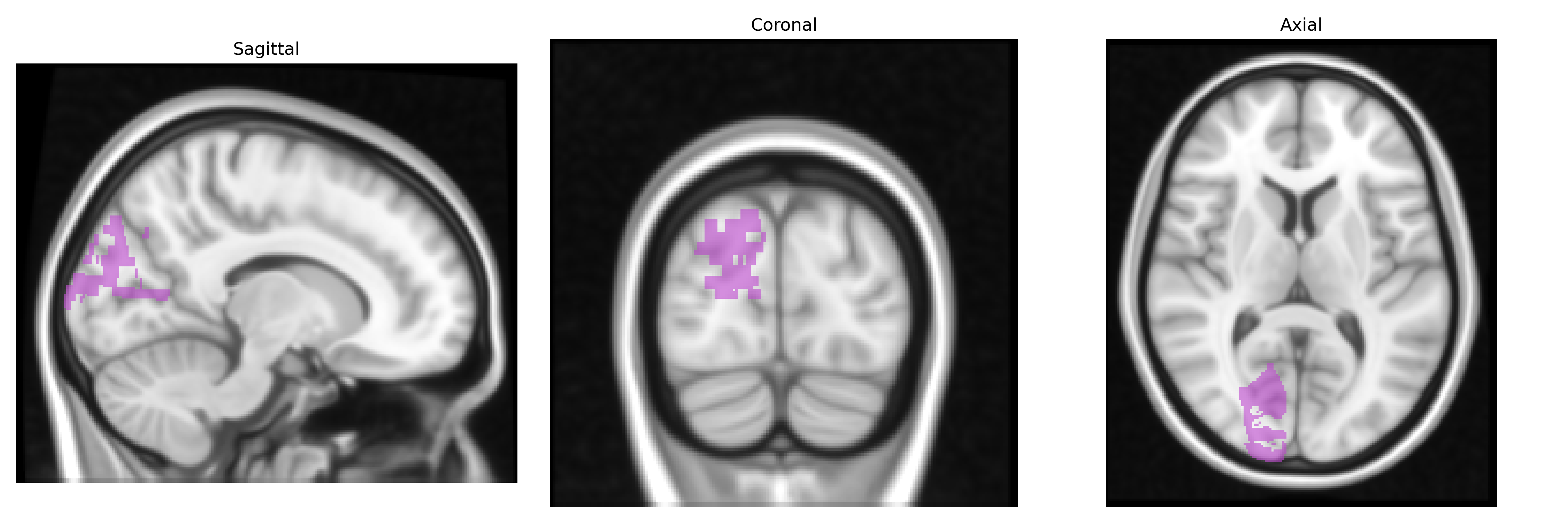
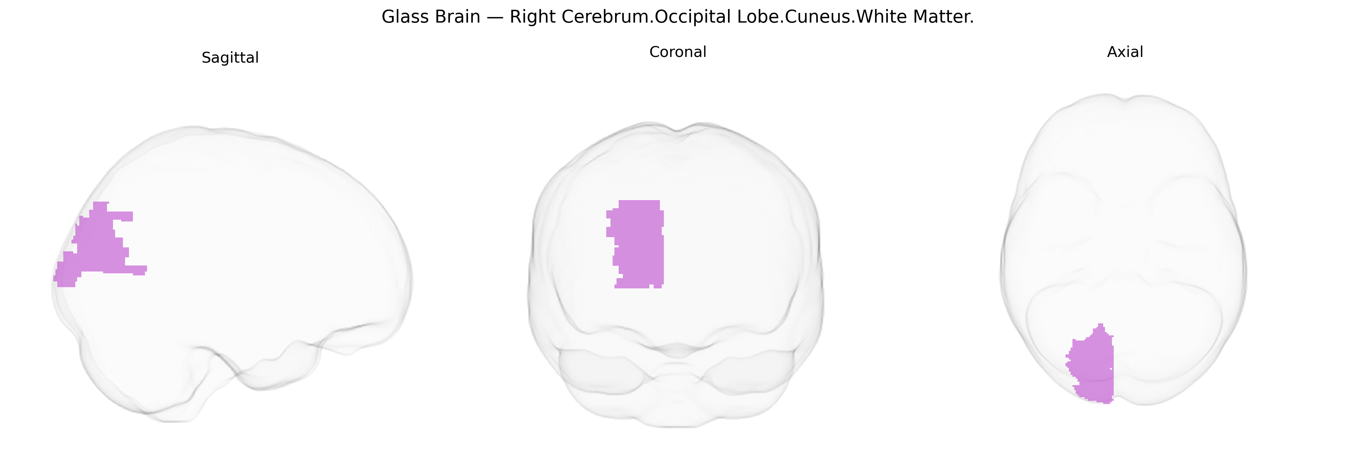
The bilateral Right Cerebrum.Occipital Lobe.Cuneus.White Matter corresponds to the deep myelinated fiber tracts underlying the cuneus region of the occipital lobe in the right cerebral hemisphere. These white matter pathways lie beneath the cortical mantle of the cuneus, a wedge-shaped area on the medial surface of the occipital lobe located superior to the calcarine sulcus and posterior to the parieto‑occipital sulcus. The fibers in this region participate in transmitting visual information between primary and associative visual cortices and other cortical and subcortical regions, contributing to processing of central visual fields, visual attention, and higher-order visuospatial functions. This white matter forms part of broader occipital and occipito-parietal connectional systems, including components of the optic radiations and association pathways interlinking dorsal visual stream areas. There is no direct Wikipedia article for this specific white matter label; a related cortical region is the Cuneus.
The bilateral right cerebrum occipital lobe cuneus white matter (Talairach 1 mm atlas) lies within posterior visual pathways and has been implicated in several imaging genetics and GWAS findings, though typically as part of broader occipital or visual network phenotypes rather than as a uniquely isolated locus. Large-scale brain MRI GWAS (e.g., UK Biobank–based studies) have identified common variants in genes related to myelination and axon guidance (such as variants near or within genes like NRG1, MAG, and PLP1) that associate with occipital white matter volume, microstructure (e.g., fractional anisotropy, mean diffusivity), or overall visual pathway integrity, which includes tracts coursing through the cuneus region. Polygenic risk for schizophrenia, bipolar disorder, and major depressive disorder has been linked to alterations in occipital white matter metrics, suggesting shared genetic architecture between psychiatric risk loci (e.g., in CACNA1C, ZNF804A, and other synaptic/neuronal genes) and posterior white matter organization. Occipital white matter, including cuneus-adjacent tracts, has also been implicated in GWAS of visual cortical thickness and surface area, where variants in neurodevelopmental genes (e.g., those involved in Wnt signaling and neuronal migration) influence structural variation across occipital regions. Additionally, genes associated with demyelinating and neurodegenerative diseases (such as HLA-DRB1 and other immune-related loci in multiple sclerosis, or APOE in Alzheimer’s disease) show associations with white matter lesion burden and integrity that include posterior periventricular and occipital territories overlapping the cuneus white matter, linking genetic susceptibility to white matter pathology and visual or cognitive impairments mediated by this region.
Overview generated by GPT-4o (2026).
Region ID: 486
Hemisphere: bilateral
Atlas: Talairach labels 1mm
Full Quality Version: Download MP4
Full Quality Version: Download MP4
Wali Sidiqyar*, Gaurav Rudravaram*, Elyssa M. McMaster, Trent M. Schwartz, Adam M. Saunders, Kurt G. Schilling, Bennett A. Landman "Introducing SPINS: A Shared Public Visualization Library of Neuroanatomical Structures." Medical Imaging with Deep Learning- short paper
This resource is licensed under CC0 1.0 Universal (Public Domain).