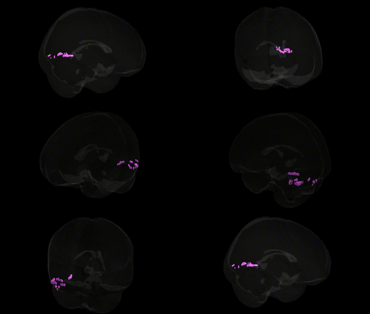
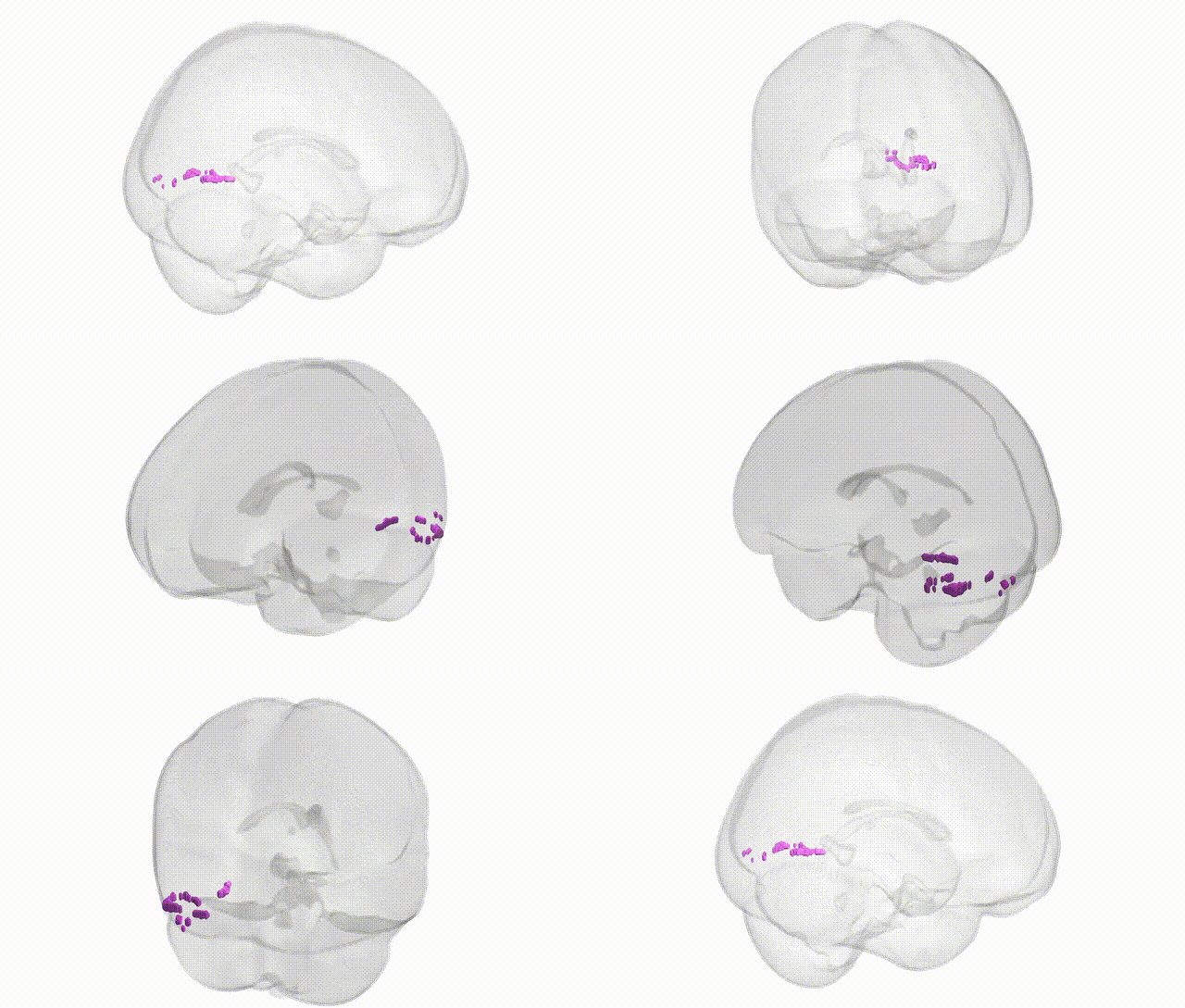
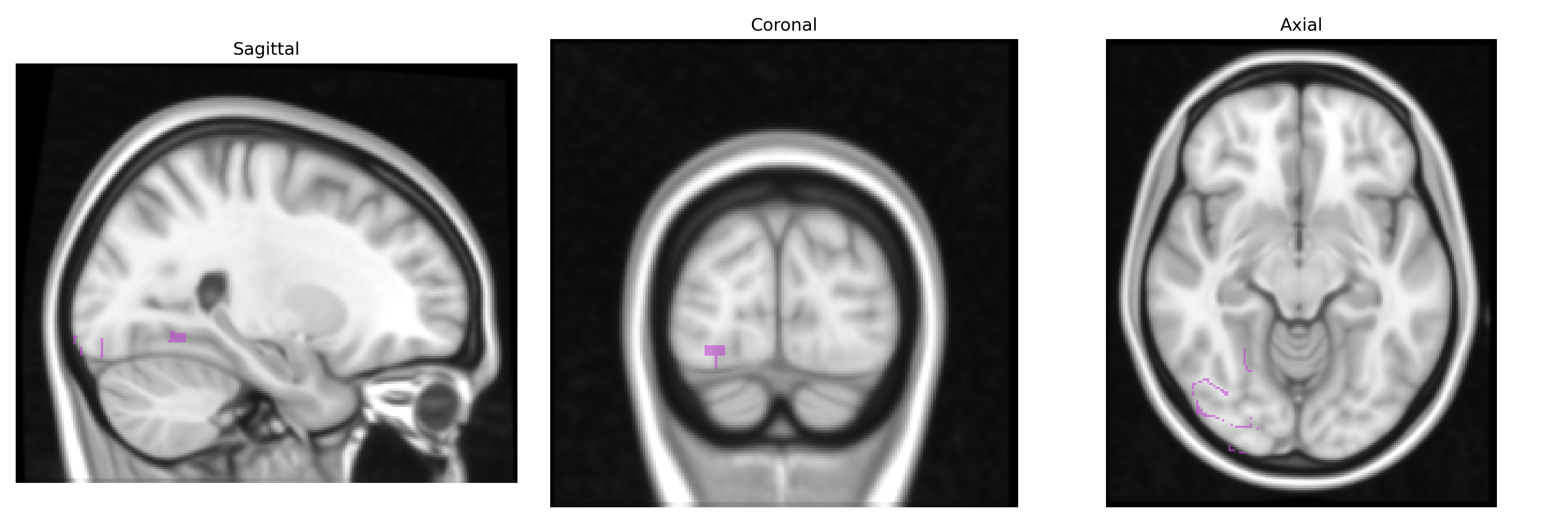
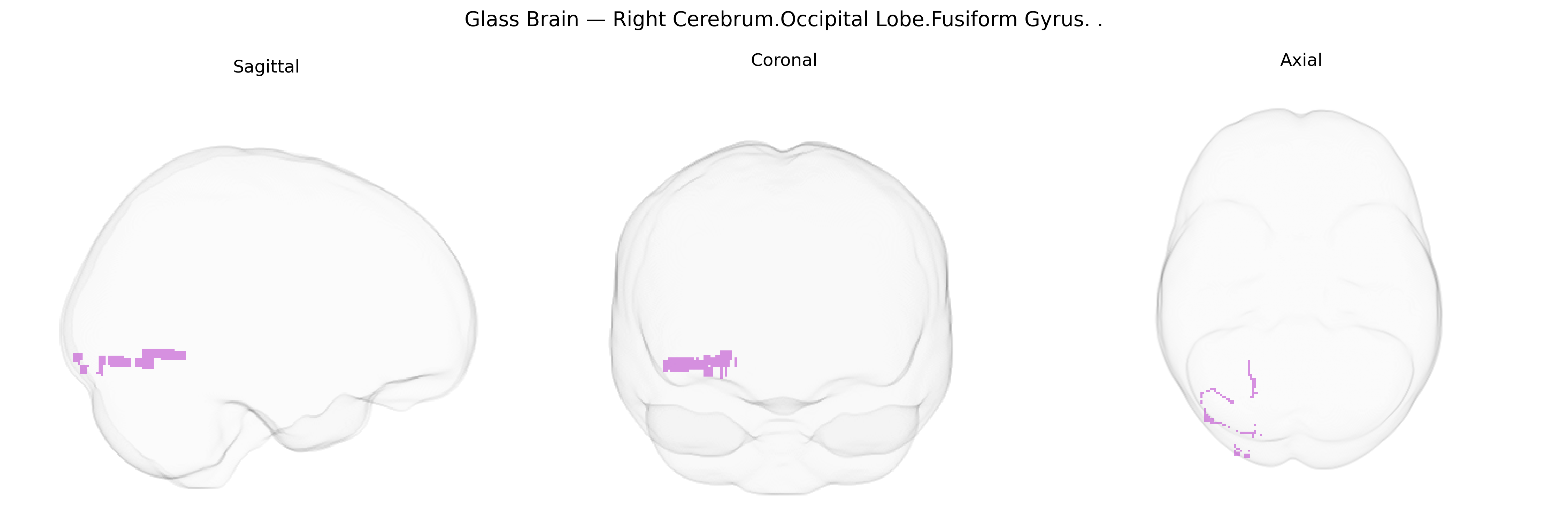
The bilateral Right Cerebrum.Occipital Lobe.Fusiform Gyrus corresponds to cortical territory within the fusiform gyrus located in the occipitotemporal region of the right hemisphere, extending along the ventral surface between the inferior temporal gyrus and the lingual gyrus. This region participates in high-level visual processing, including complex pattern and object recognition, and contributes to category-selective representations such as faces, words, and other visually defined classes, often in concert with homologous left-hemisphere areas. Its cytoarchitecture is part of the ventral visual stream, receiving processed visual input from occipital visual areas and projecting to temporal and limbic regions involved in memory and semantic integration. Although the Talairach label emphasizes occipital lobe localization, functionally the fusiform gyrus is considered part of the occipitotemporal cortex and is implicated in perceptual expertise and visual identification. Fusiform gyrus
The bilateral right cerebrum occipital fusiform gyrus—overlapping with what many neuroimaging genetics studies call the fusiform gyrus, occipitotemporal cortex, or visual word form/face-selective regions—has been implicated in several genetic and GWAS-derived associations, primarily via imaging genetics rather than direct Talairach-based studies. Twin and family studies show high heritability for fusiform/occipitotemporal cortical thickness and surface area, with specific loci (e.g., near HMGA2, IGF1, and other growth or neurodevelopmental genes) associated with occipital and ventral temporal morphology in large consortia such as ENIGMA and UK Biobank–based GWAS. Genetic variants in or near KIAA0319, DCDC2, ROBO1, and other dyslexia-susceptibility genes have been linked to altered structure and activation in left and bilateral fusiform/occipitotemporal regions during reading, while face-processing endophenotypes and fusiform morphology show heritable components and candidate-gene associations (including regions near genes involved in synaptic development). Autism spectrum disorder, developmental prosopagnosia, and schizophrenia exhibit both structural and functional abnormalities in fusiform and occipital fusiform cortex, with polygenic risk scores and common variants in synaptic, neurodevelopmental, and chromatin-remodeling genes (e.g., SHANK3, NRXN1, CNTNAP2, and others) correlating with fusiform/occipital volume or activation differences in some cohorts. GWAS of visual cortical thickness and occipital lobe volume also identify multiple loci involved in neural proliferation, axon guidance, and myelination, suggesting that genetic architecture of the fusiform/occipital region is polygenic and overlaps with risk pathways for neurodevelopmental disorders, learning disabilities, and variation in higher-order visual and reading abilities.
Overview generated by GPT-4o (2026).
Region ID: 204
Hemisphere: bilateral
Atlas: Talairach labels 1mm
Full Quality Version: Download MP4
Full Quality Version: Download MP4
Wali Sidiqyar*, Gaurav Rudravaram*, Elyssa M. McMaster, Trent M. Schwartz, Adam M. Saunders, Kurt G. Schilling, Bennett A. Landman "Introducing SPINS: A Shared Public Visualization Library of Neuroanatomical Structures." Medical Imaging with Deep Learning- short paper
This resource is licensed under CC0 1.0 Universal (Public Domain).