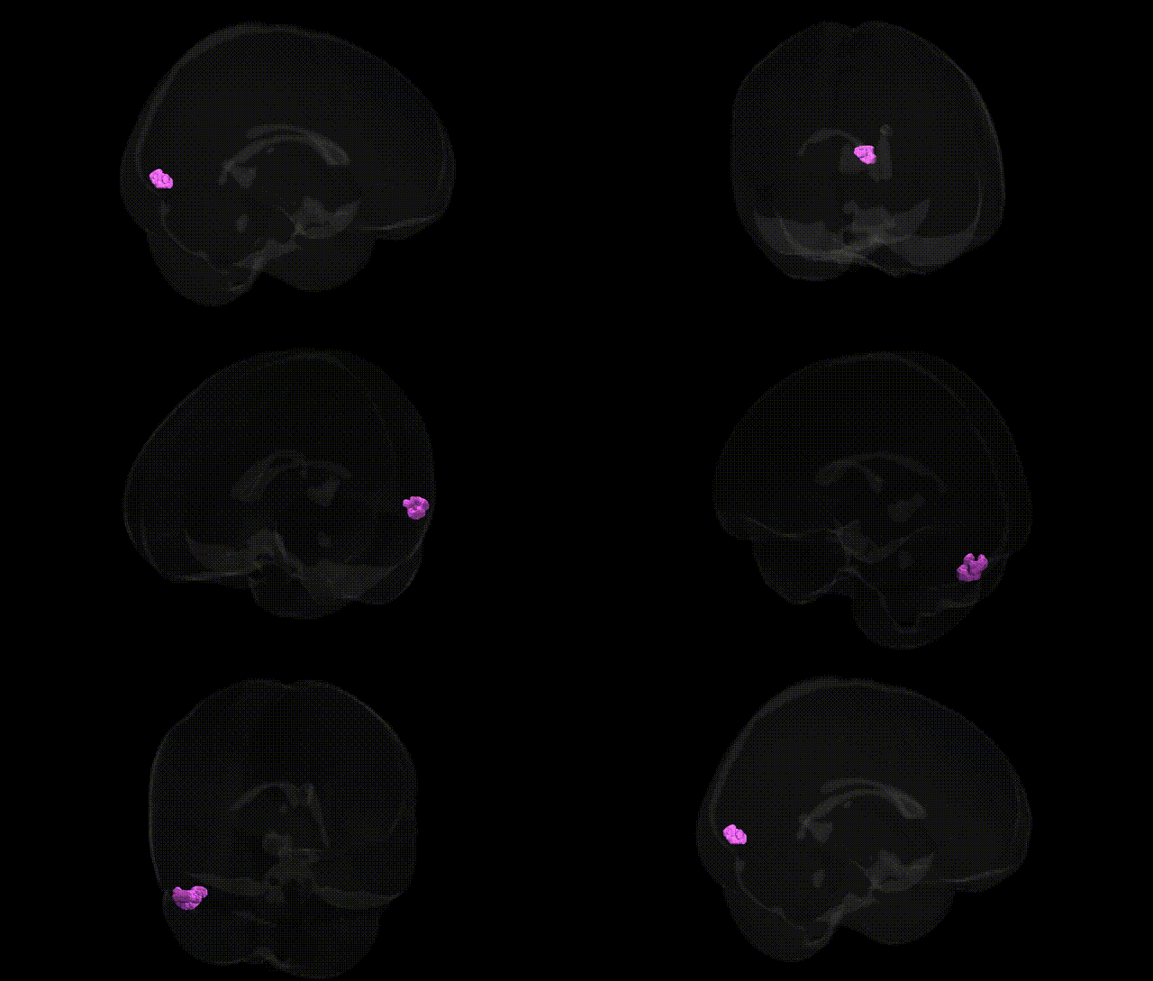
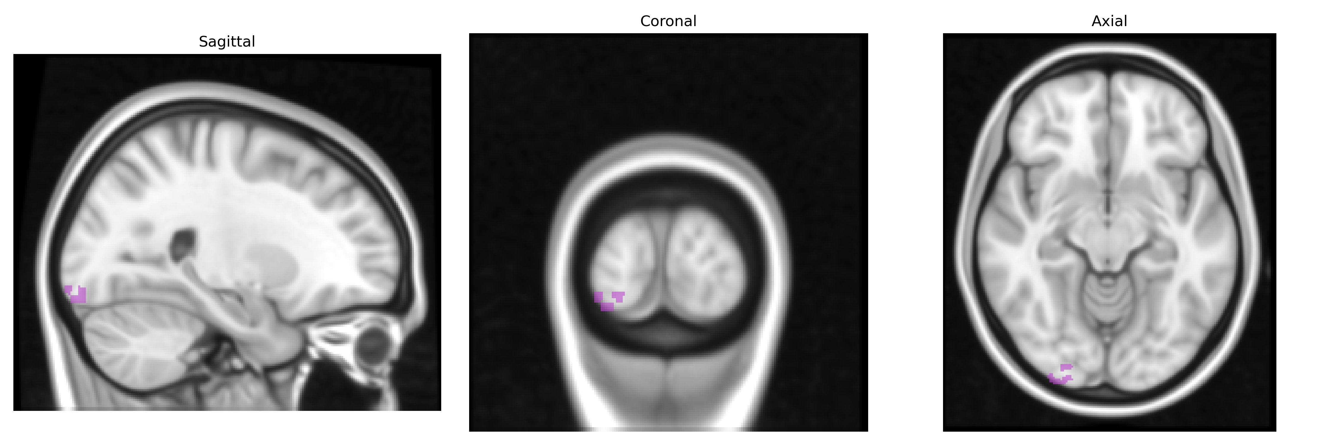
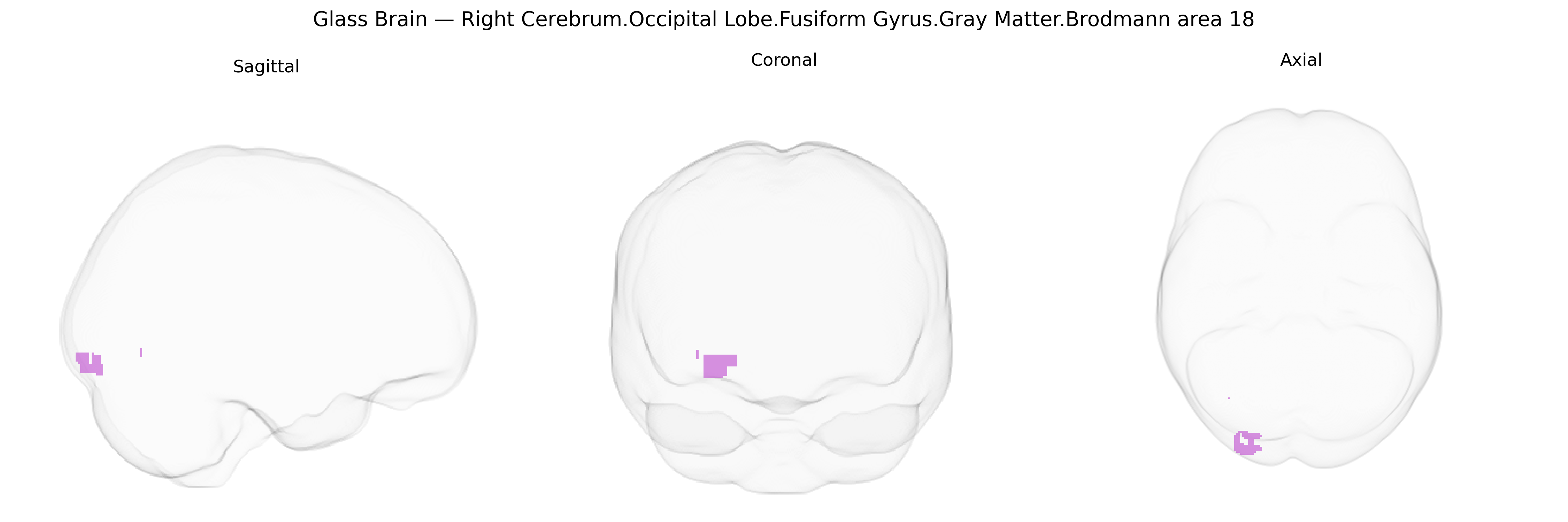
The bilateral Right Cerebrum.Occipital Lobe.Fusiform Gyrus.Gray Matter.Brodmann area 18 corresponds to secondary visual association cortex located along the fusiform gyrus within the occipital lobe, adjacent to primary visual cortex (Brodmann area 17). This region participates in higher-order processing of visual information, including complex pattern, form, and object recognition, and serves as an intermediate stage between early visual input and more specialized visual areas involved in category-specific processing (such as faces and words). Cytoarchitectonically, it is characterized by a well-developed granular layer IV and distinct lamination, reflecting its role in receiving dense input from primary visual cortex and distributing processed signals to temporal and parietal association areas. There is no direct link for this exact composite label; a related structure is Brodmann area 18.
Genetic associations specifically targeting bilateral Right Cerebrum.Occipital Lobe.Fusiform Gyrus.Gray Matter.Brodmann area 18 (BA18) are typically inferred from broader studies of occipital cortex, fusiform gyrus, or visual association areas, as most GWAS use macro-level parcellations rather than Talairach-based BA18 labels at 1 mm resolution. BA18, a secondary visual (association) cortex, overlaps with regions implicated in visual perception, face and word recognition, and high-level object processing; GWAS of cortical thickness and surface area have implicated variants near genes involved in neurodevelopment, synaptic function, and axon guidance (e.g., MIR2113, HMGA2, and other loci reported in ENIGMA and UK Biobank-based imaging genetics studies) that show effects in occipital and ventral temporal regions, including fusiform/visual association cortex. Genetic studies of functional activation and structural variation in fusiform/occipital association areas implicate risk loci for neurodevelopmental and neuropsychiatric conditions, such as autism spectrum disorder, schizophrenia, and dyslexia, in which altered fusiform and occipital volumes or activation patterns are observed; candidate and GWAS-identified genes related to synaptic plasticity and cortical patterning (e.g., NRGN, CNTNAP2, and others) have been linked to atypical structure–function coupling in these regions. GWAS of face recognition ability and prosopagnosia have reported heritable contributions and associations with variants near genes involved in visual and social brain network development, consistent with the known role of fusiform/occipital association cortex in face processing. In addition, large-scale imaging-genetics consortia have found that polygenic scores for educational attainment, intelligence, and neuroticism correlate with differences in occipital and fusiform cortical measures that include BA18 territory, suggesting that distributed genetic influences on cognition and affect partially manifest in structural and functional variation of this visual association region, although precise BA18-specific loci remain incompletely resolved and are typically inferred from overlapping atlases rather than directly annotated to the Talairach labels 1 mm Atlas region.
Overview generated by GPT-4o (2026).
Region ID: 205
Hemisphere: bilateral
Atlas: Talairach labels 1mm
Full Quality Version: Download MP4
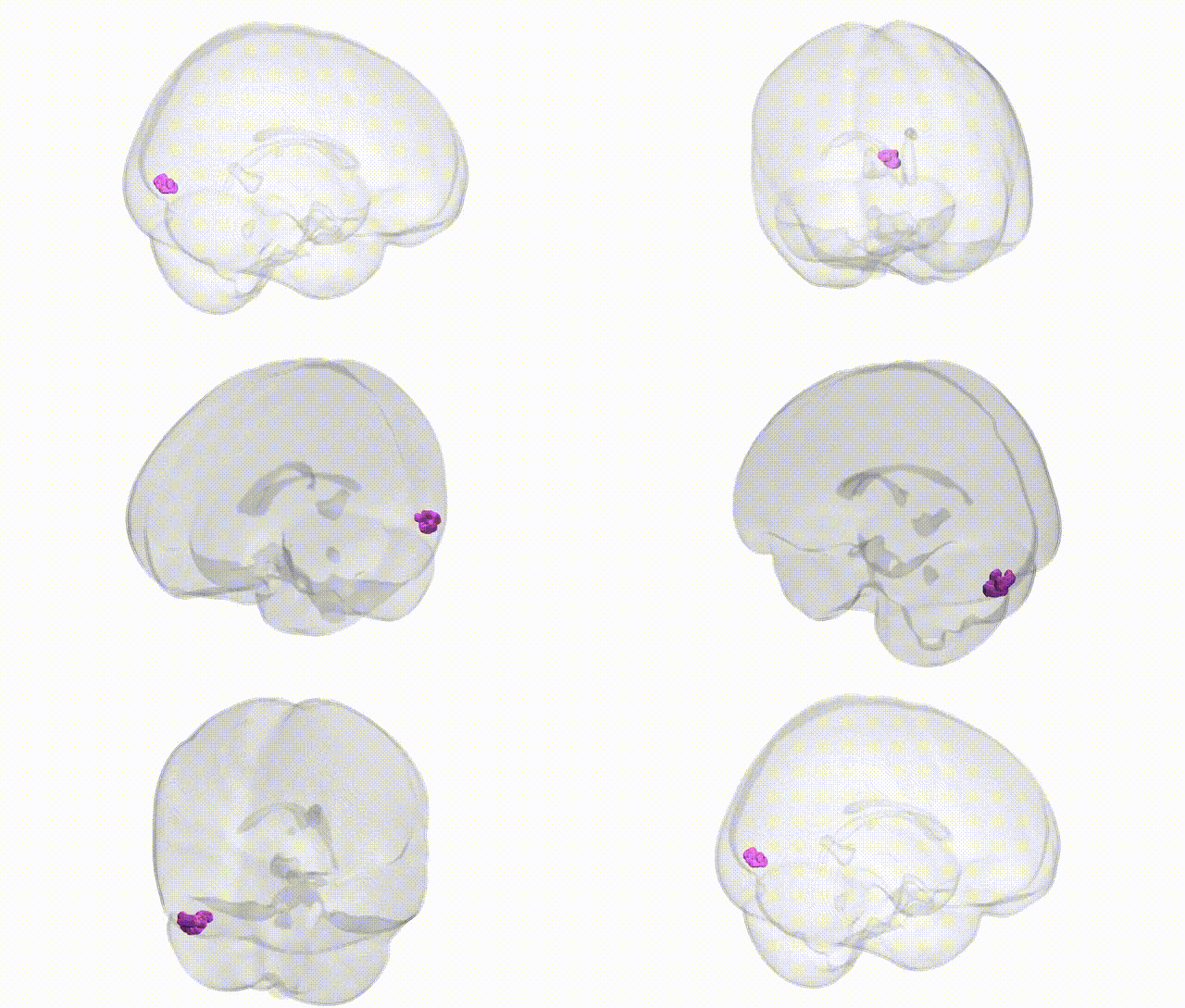
Full Quality Version: Download MP4
Wali Sidiqyar*, Gaurav Rudravaram*, Elyssa M. McMaster, Trent M. Schwartz, Adam M. Saunders, Kurt G. Schilling, Bennett A. Landman "Introducing SPINS: A Shared Public Visualization Library of Neuroanatomical Structures." Medical Imaging with Deep Learning- short paper
This resource is licensed under CC0 1.0 Universal (Public Domain).