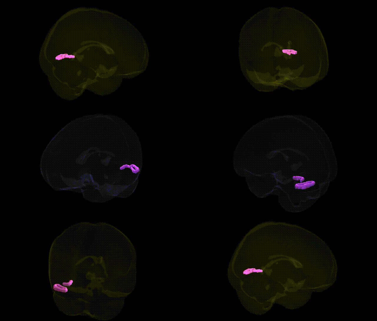
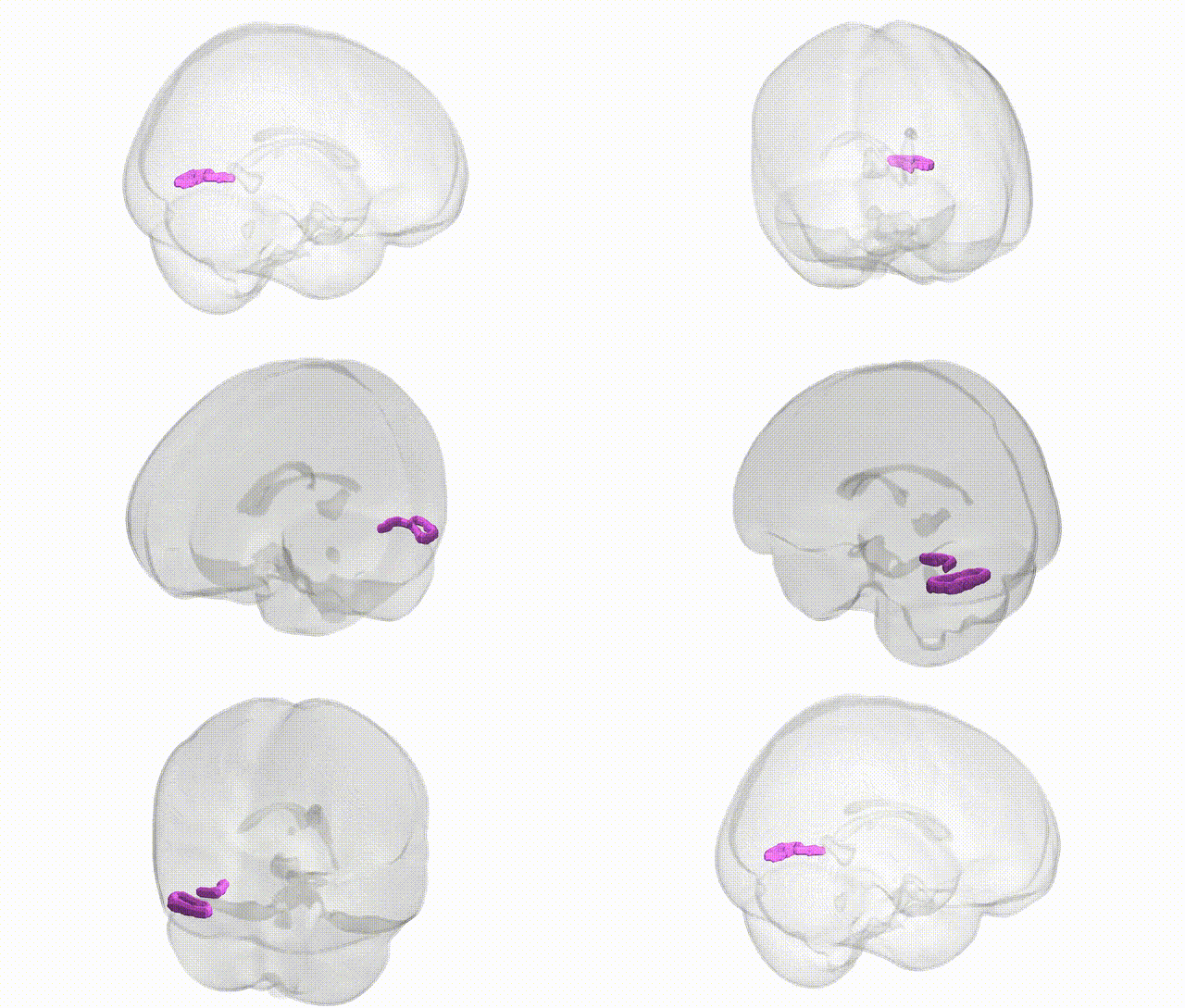
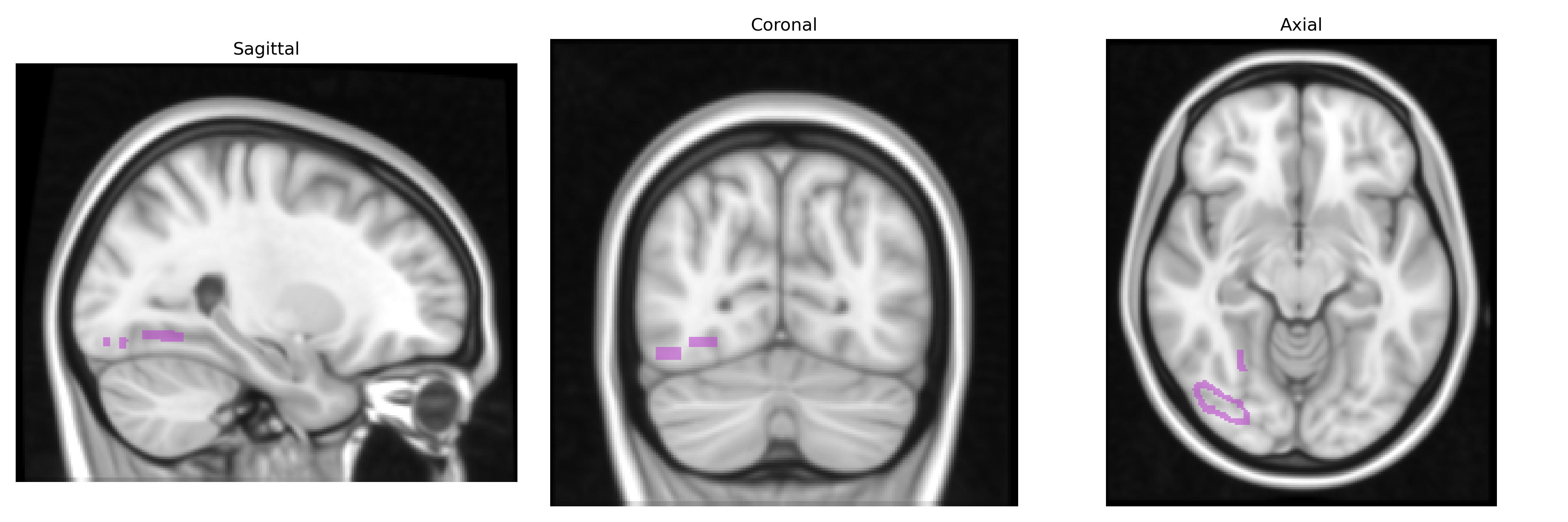
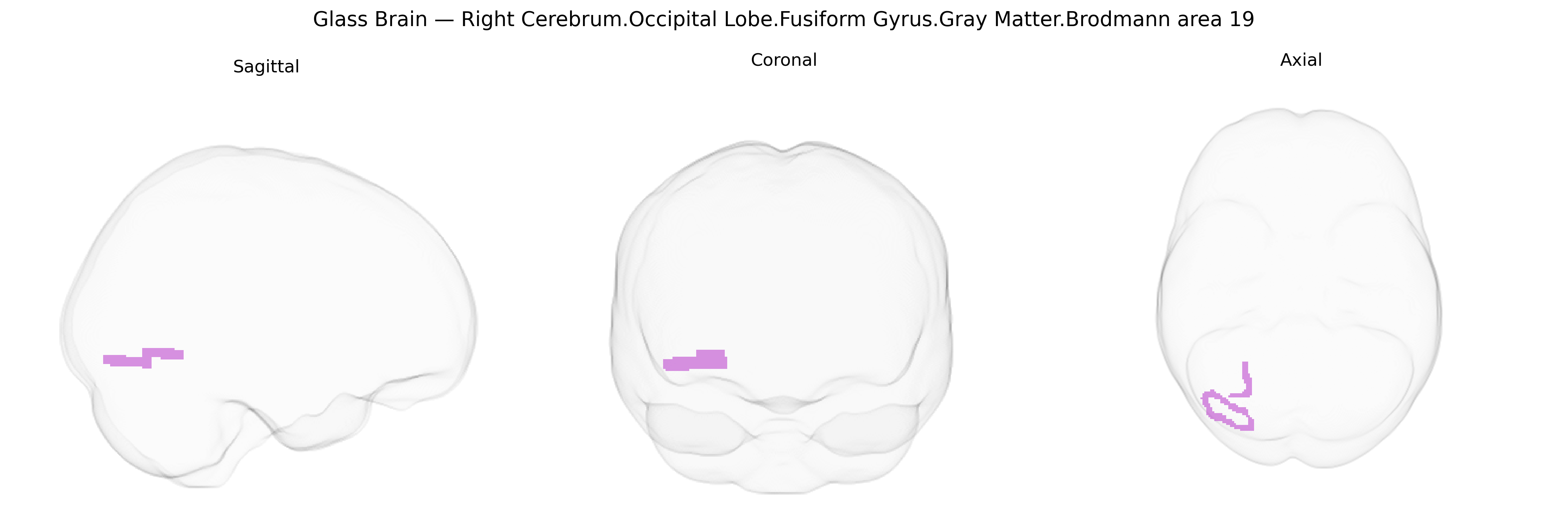
The bilateral Right Cerebrum.Occipital Lobe.Fusiform Gyrus.Gray Matter.Brodmann area 19 corresponds to a higher-order visual association region located in the posterior fusiform gyrus of the occipital lobe, within the right cerebral hemisphere. As part of Brodmann area 19, it participates in complex visual processing, including integration of contours, motion, color, and form, and contributes to object, face, and scene recognition by relaying and transforming information from primary and secondary visual cortices. Neuronal populations in this gray matter region exhibit modality-specific and multimodal responses, supporting the abstraction of visual features into coherent percepts. Functionally, it is embedded in the ventral visual stream (“what” pathway), interacting with adjacent fusiform and occipitotemporal territories involved in category-selective recognition. There is no direct Wikipedia article for this precise Talairach label; a closely related structure is the Fusiform gyrus.
The bilateral right occipital fusiform gyrus gray matter within Brodmann area 19, as defined by the Talairach 1 mm atlas, is part of higher-order visual association cortex and has been implicated in several genetic and GWAS-derived associations, though typically at the level of broader occipitotemporal or fusiform regions rather than this exact microanatomical label. Large-scale brain imaging GWAS (e.g., ENIGMA and UK Biobank consortia) have identified common variants in genes involved in neurodevelopment, axon guidance, and synaptic signaling (such as variants near HMGA2, MAPT, and various cell adhesion and axon guidance genes) that influence occipital and fusiform cortical thickness, surface area, and volume, with some loci showing specificity for visual association regions including BA18/19. The fusiform/occipital complex has also been tied to genetic liability for neurodevelopmental and psychiatric disorders: polygenic risk for autism spectrum disorder, schizophrenia, and major depression has been associated with structural and functional alterations in fusiform and occipital association cortices, and rare copy number variants (e.g., 22q11.2 deletions) affecting neurodevelopmental pathways are linked to aberrant fusiform and visual association morphology. In addition, GWAS of face recognition ability and reading-related traits (including dyslexia) have implicated genes involved in cortical patterning and synaptic plasticity that modulate activity and structure in the fusiform/occipitotemporal “visual word form area” and face-selective cortex, which overlap with BA19 territory. However, most genetic findings remain regionally coarse and are not specific to the Talairach-defined “Right Cerebrum.Occipital Lobe.Fusiform Gyrus.Gray Matter.Brodmann area 19” label, so current evidence is best viewed as implicating this region as part of a broader genetically influenced occipitotemporal network supporting high-level visual processing and associated cognitive traits.
Overview generated by GPT-4o (2026).
Region ID: 259
Hemisphere: bilateral
Atlas: Talairach labels 1mm
Full Quality Version: Download MP4
Full Quality Version: Download MP4
Wali Sidiqyar*, Gaurav Rudravaram*, Elyssa M. McMaster, Trent M. Schwartz, Adam M. Saunders, Kurt G. Schilling, Bennett A. Landman "Introducing SPINS: A Shared Public Visualization Library of Neuroanatomical Structures." Medical Imaging with Deep Learning- short paper
This resource is licensed under CC0 1.0 Universal (Public Domain).