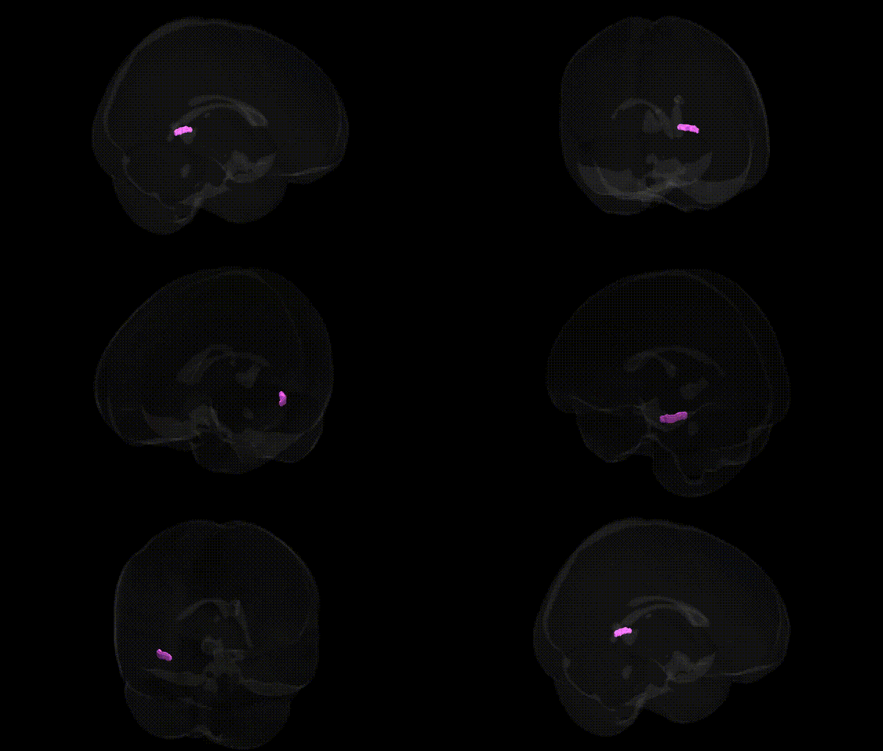
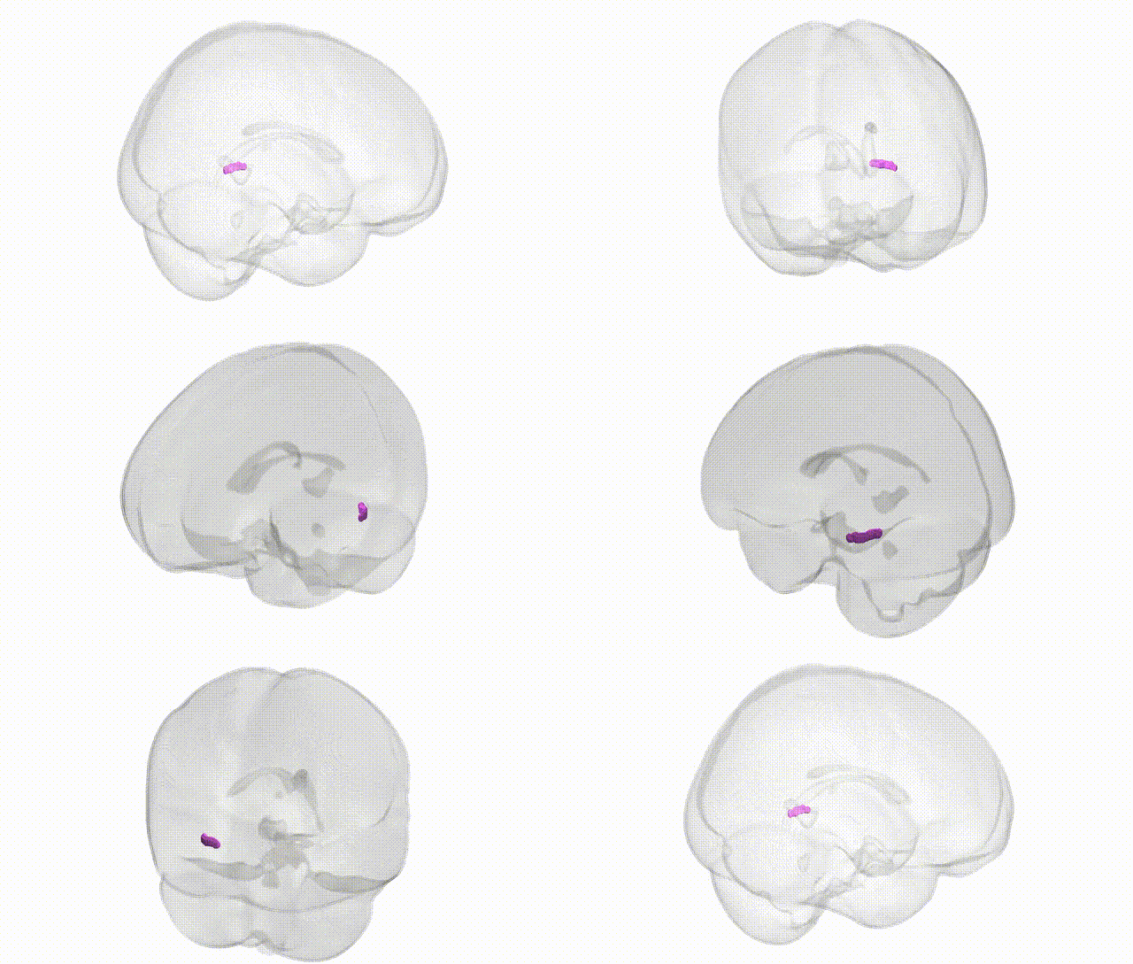
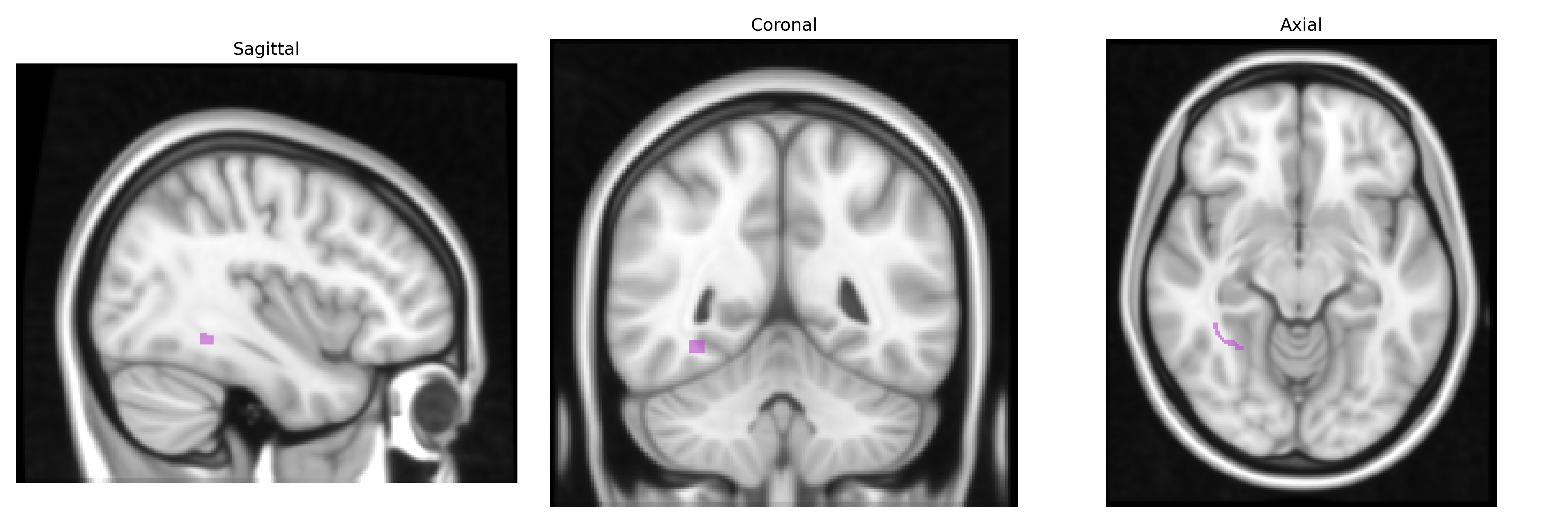
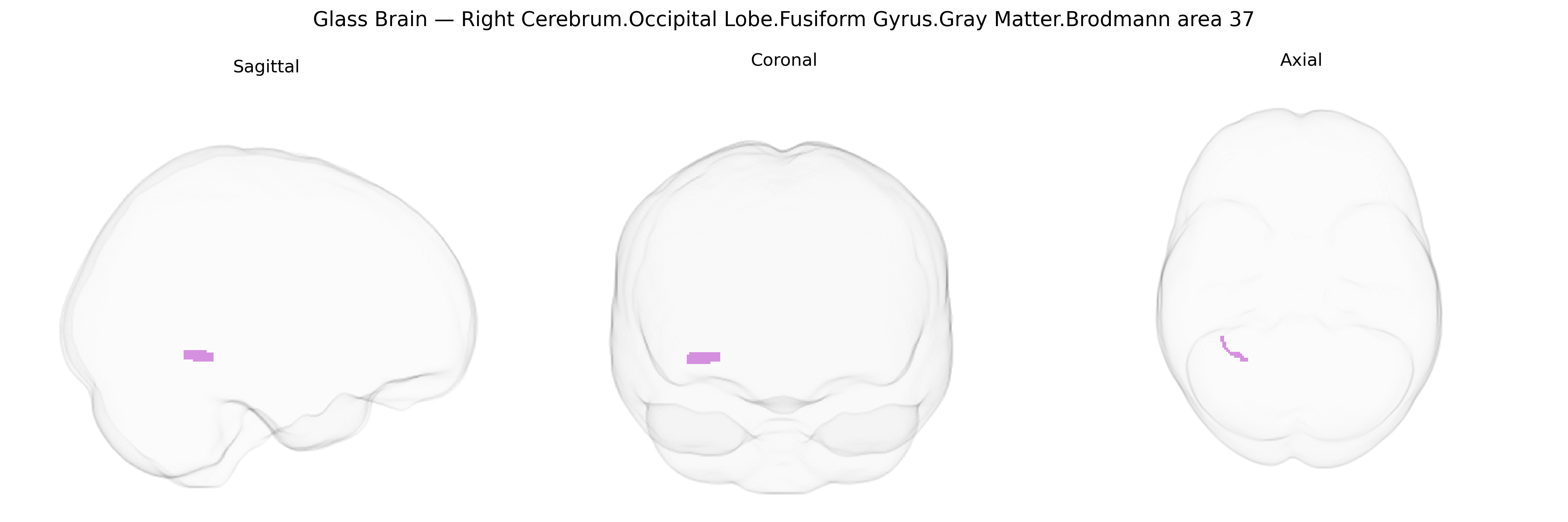
The bilateral Right Cerebrum.Occipital Lobe.Fusiform Gyrus.Gray Matter.Brodmann area 37 corresponds to a cytoarchitectonic region located in the posterior-inferior temporal–occipital cortex, primarily within the fusiform gyrus and adjacent portions of the inferior temporal and occipital lobes. Brodmann area 37 is associated with high-level visual processing, including object recognition, word-form processing, and aspects of face and category-specific perception, integrating complex visual inputs with stored semantic representations. It receives afferent projections from earlier visual areas and projects to temporal and frontal association cortices, contributing to the ventral “what” visual processing stream and playing a role in multimodal integration and language-related functions. There is no direct link for “Brodmann area 37” as a standalone article; a closely related structure is the fusiform gyrus: Fusiform gyrus.
The bilateral right fusiform gyrus in Brodmann area 37, part of the occipitotemporal cortex, has been repeatedly implicated in genetic and GWAS-based studies of face and word recognition, social cognition, and neurodevelopmental and psychiatric conditions. Twin and family studies show high heritability of fusiform gyrus volume and functional responses, with common variant GWAS linking structural variation in this region to loci involving neuronal development, synaptic function, and axonal guidance (for example, variants near genes such as DLG2, PAX6, and CNTNAP2 in some imaging-genetics cohorts), although specific effects often span broader occipitotemporal networks rather than BA37 alone. In neurodevelopmental disorders, risk variants for autism spectrum disorder, dyslexia, and specific language impairment overlap with genes whose expression and downstream effects involve fusiform and adjacent temporal regions, consistent with altered fusiform structure and activation observed in these conditions. In schizophrenia and bipolar disorder, polygenic risk scores and GWAS hits for synaptic and glutamatergic genes have been associated with reduced fusiform gray matter volume and disrupted connectivity, potentially contributing to deficits in face processing and visual memory. GWAS of face recognition ability and social perception traits also show that individual differences in fusiform activation and morphology are partially explained by common genetic variation, including loci in or near genes related to neurodevelopmental patterning and cortical column organization, supporting a genetically influenced specialization of the right BA37 fusiform region for complex visual object and face processing.
Overview generated by GPT-4o (2026).
Region ID: 330
Hemisphere: bilateral
Atlas: Talairach labels 1mm
Full Quality Version: Download MP4
Full Quality Version: Download MP4
Wali Sidiqyar*, Gaurav Rudravaram*, Elyssa M. McMaster, Trent M. Schwartz, Adam M. Saunders, Kurt G. Schilling, Bennett A. Landman "Introducing SPINS: A Shared Public Visualization Library of Neuroanatomical Structures." Medical Imaging with Deep Learning- short paper
This resource is licensed under CC0 1.0 Universal (Public Domain).